Review
. 2007 Oct 15;74(8):1235-46.
doi: 10.1016/j.bcp.2007.07.032. Epub 2007 Jul 27.

The subtypes of nicotinic acetylcholine receptors on dopaminergic terminals of mouse striatum

Affiliations
Review

The subtypes of nicotinic acetylcholine receptors on dopaminergic terminals of mouse striatum

Sharon R Grady et al. Biochem Pharmacol. .

Abstract

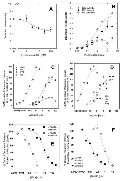
This review summarizes studies that attempted to determine the subtypes of nicotinic acetylcholine receptors (nAChR) expressed in the dopaminergic nerve terminals in the mouse. A variety of experimental approaches has been necessary to reach current knowledge of these subtypes, including in situ hybridization, agonist and antagonist binding, function measured by neurotransmitter release from synaptosomal preparations, and immunoprecipitation by selective antibodies. Early developments that facilitated this effort include the radioactive labeling of selective binding agents, such as [(125)I]-alpha-bungarotoxin and [(3)H]-nicotine, advances in cloning the subunits, and expression and evaluation of function of combinations of subunits in Xenopus oocytes. The discovery of epibatidine and alpha-conotoxin MII (alpha-CtxMII), and the development of nAChR subunit null mutant mice have been invaluable in determining which nAChR subunits are important for expression and function in mice, as well as allowing validation of the specificity of subunit specific antibodies. These approaches have identified five nAChR subtypes of nAChR that are expressed on dopaminergic nerve terminals. Three of these contain the alpha6 subunit (alpha4alpha6beta2beta3, alpha6beta2beta3, alpha6beta2) and bind alpha-CtxMII with high affinity. One of these three subtypes (alpha4alpha6beta2beta3) also has the highest sensitivity to nicotine of any native nAChR that has been studied, to date. The two subtypes that do not have high affinity for alpha-CtxMII (alpha4beta2, alpha4alpha5beta2) are somewhat more numerous than the alpha6* subtypes, but do bind nicotine with high affinity. Given that our first studies detected readily measured differences in sensitivity to agonists and antagonists among these five nAChR subtypes, it seems likely that subtype selective compounds could be developed that would allow therapeutic manipulation of diverse nAChRs that have been implicated in a number of human conditions.


Figures

Figure 1
Figure 1
in situ at level of SN/VTA. Frozen sections (14μm) from a C57Bl6 mouse at the level of SN/VTA (∼-3.0mm bregma) were hybridized with full length riboprobes complementary to the α2, α3, α4, α5, α6, α7, β2, β3 and β4 nAChR subunits [64,100].
Figure 2
Figure 2
nAChR binding sites measurable with radiolabeled epibatidine. This diagram illustrates total mouse brain radiolabeled-epibatidine binding and the methods that can be used to subdivide this binding. Epibatidine binding in mouse brain is biphasic, with two sites differing significantly in affinity. Higher affinity refers to those receptor subtypes with KD values of ∼ 0.02 nM, while lower affinity receptors have KD values of ∼ 5 nM. Percentages throughout the diagram indicate the relative abundance of particular subsets in whole brain; however, note that relative abundance does vary among brain regions. “Required” subunits are determined by loss of binding in null mutant mice. Partial dependence of binding on a subunit is noted by “sometimes”, “significant” or “primarily”. Two of these subsets have been well-characterized by other methods. Namely, higher-affinity cytisine-sensitive binding is the subset that is also measured directly by [3H]-nicotine or [3H]-cytisine binding, while the lower-affinity αBgt-sensitive subset is the nAChR measured directly by [125I]-α-bungarotoxin binding. In addition, the α-CtxMII-sensitive subset can be measured directly by [125I]-α-CtxMII binding (Ki <1nM). The α-CtxMII-resistant subset includes some receptors with moderate affinity for α-CtxMII (Ki >10nM).
Figure 3
Figure 3
nAChR binding in striatum. Specific binding to nAChRs was measured in striatal membrane preparations from mice of the three genotypes (wildtype, heterozygous, and null mutant) of each nAChR subunit null mutation indicated on the x-axis. Panel A: Binding representing the α4β2* subtypes was measured by nicotine binding for the β3 null genotypes [100], and by higher-affinity cytisine-sensitive [3H]epibatidine binding for the remaining genotypes (α5 data from [86]; β2 from [119]; α4, α7 and β4 unpublished data from MJ Marks). Panel B: Binding representing the α6β2* subtypes was measured by [125I]-α-CtxMII binding (from [86]). For both panels, significant differences (1-way ANOVA) are indicated by * (different from wildtype P<0.05) or + (different from heterozygote P<0.05).
Figure 4
Figure 4
Agonist-stimulated dopamine release from striatal synaptosomes. Panel A: α-CtxMII partially inhibits nicotine-stimulated dopamine release, evoked by a 10s exposure to 10 μM ACh, with an IC50 value of 2.2nM (data from [63]). Units indicate dopamine release as cpm normalized to cpm of baseline release. Panel B: ACh-stimulated dopamine release measured from C57Bl striatal synaptosomes with (MII resistant activity) and without (total activity) prior exposure to α-CtxMII (50nM for 5 min). The MII-sensitive activity is determined by difference (from [42]). Units are calculated as for Panel A. Panel C: α-CtxMII-resistant dopamine release stimulated by various agonists as % of maximal α-CtxMII-resistant release by ACh (replotted from [42]). Panel D: α-CtxMII-sensitive dopamine release stimulated by various agonists as % of maximal α-CtxMII-sensitive release by ACh (replotted from [42]). Panel E: Inhibition of both α-CtxMII-resistant and —sensitive dopamine release stimulated by nicotine (3μM) by MLA (from [42]). Panel F: Inhibition of both α-CtxMII-resistant and —sensitive dopamine release stimulated by nicotine (3μM) by DHβE (from [42]).
Figure 5
Figure 5
Effect of nAChR subunit null mutations on agonist-stimulated dopamine release from striatal synaptosomes. Panel A: Effects on α-CtxMII-resistant dopamine release by ACh (10μM) with prior exposure to α-CtxMII (50nM for 5 min) as % of wildtype response (replotted from [42] and unpublished data from SR Grady). Panel B: Effects on α-CtxMII-sensitive dopamine release stimulated by ACh (1μM) by difference between samples of synaptosomes with and without prior exposure to α-CtxMII (50nM for 5 min) as % of wildtype response (replotted from [42] and unpublished data from SR Grady). For both panels significant differences (1-way ANOVA) are indicated by * (different from wildtype P<0.05) or + (different from heterozygote P<0.05).

References

    1. Martin LF, Kem WR, Freedman R. Alpha-7 nicotinic receptor agonists: potential new candidates for the treatment of schizophrenia. Psychopharmacology. 2004;174:54–64. - PubMed
    1. Singh A, Potter A, Newhouse P. Nicotinic acetylcholine receptor system and neuropsychiatric disorders. Idrugs. 2004;7:1096–103. - PubMed
    1. Potter AS, Newhouse PA, Bucci DJ. Central nicotinic cholinergic systems: a role in the cognitive dysfunction in attention-deficit/hyperactivity disorder? Behav Brain Research. 2006;175:201–11. - PubMed
    1. Steinlein OK. Nicotinic receptor mutations in human epilepsy. Prog in Brain Res. 2004;145:275–85. - PubMed
    1. Quik M, O’Neill M, Perez XA. Nicotine neuroprotection against nigrostriatal damage: importance of the animal model. Trends in Pharmacol Sci. 2007;28:229–35. - PubMed

Publication types