. 2007 Dec 20;369(2):389-99.
doi: 10.1016/j.virol.2007.07.029. Epub 2007 Sep 6.

An ATPase activity associated with the rotavirus phosphoprotein NSP5

Affiliations

An ATPase activity associated with the rotavirus phosphoprotein NSP5

Tamara Bar-Magen et al. Virology. .

Abstract

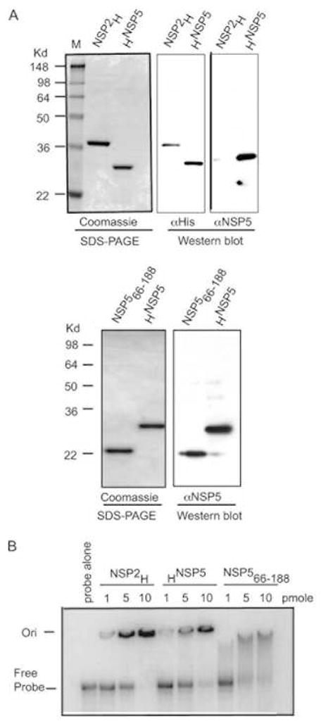
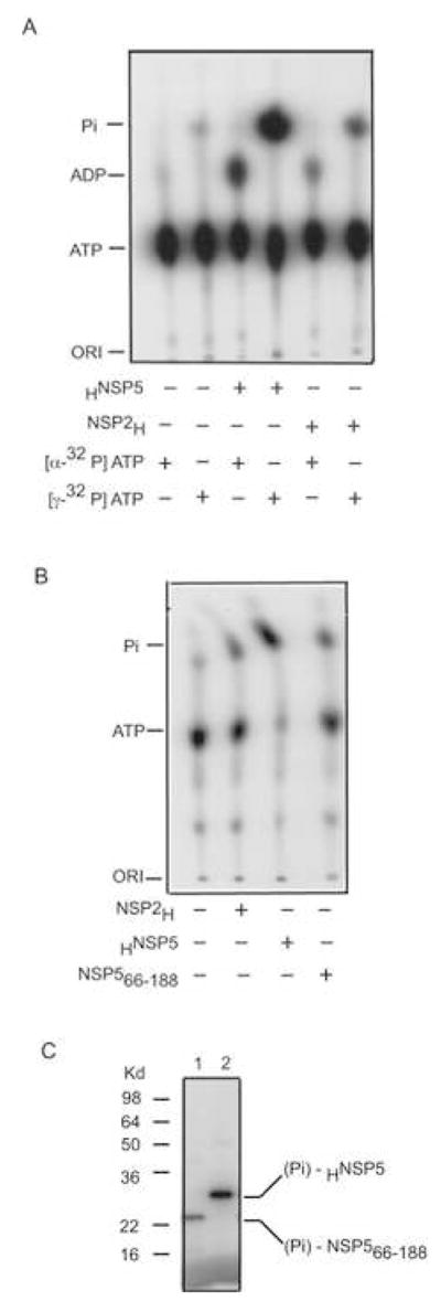
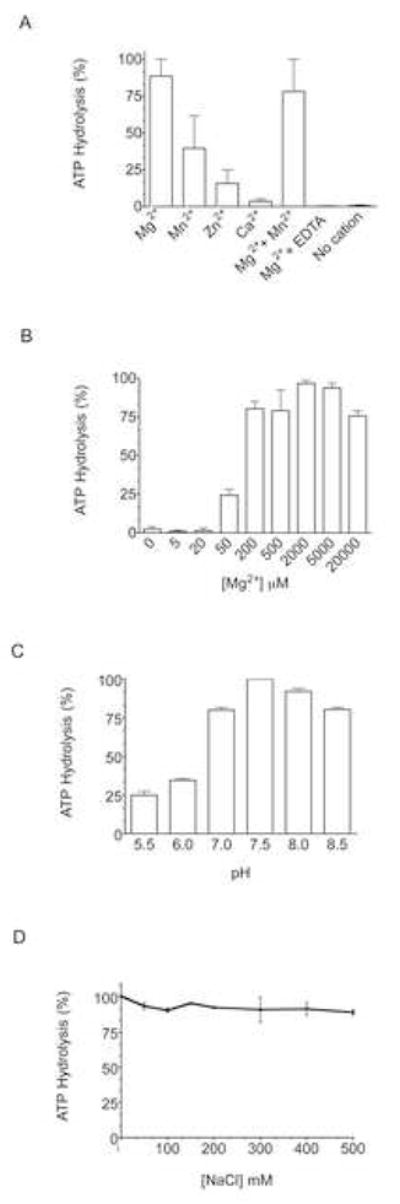
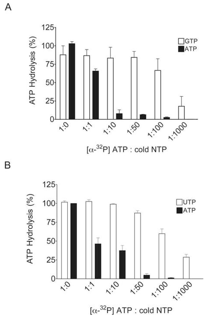
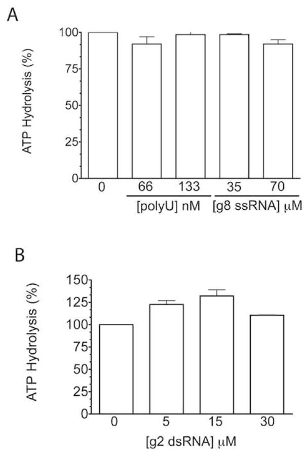
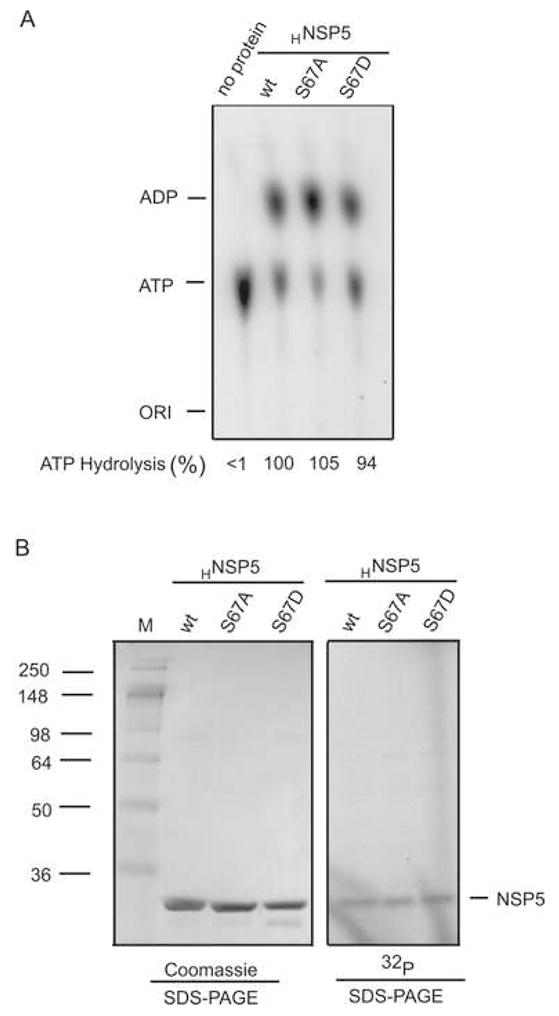
Interactions between NSP5 and NSP2 drive the formation of viroplasms, sites of genome replication and packaging in rotavirus-infected cells. The serine-threonine-rich NSP5 transitions between hypo- and hyper-phosphorylated isomers during the replication cycle. In this study, we determined that purified recombinant NSP5 has a Mg2+-dependent ATP-specific triphosphatase activity that generates free ADP and Pi (Vmax of 19.33 fmol of product/min/pmol of enzyme). The ATPase activity was correlated with low levels of NSP5 phosphorylation, suggestive of a possible link between ATP hydrolysis and an NSP5 autokinase activity. Mutagenesis showed that the critical residue (Ser67) needed for NSP5 hyperphosphorylation by cellular casein kinase-like enzymes has no role in the ATPase or autokinase activities of NSP5. Through its NDP kinase activity, the NSP2 octamer may support NSP5 phosphorylation by creating a constant source of ATP molecules for the autokinase activity of NSP5 and for cellular kinases associated with NSP5.


Figures

Figure 1
Figure 1. Purified recombinant proteins
(A) His-tagged full-length NSP2 (NSP2H) and NSP5 (HNSP5) and intein-tagged NSP566–188 were expressed in bacteria and purified by affinity chromatography. Purified proteins were resolved by polyacrylamide-gel electrophoresis (PAGE), and detected by staining with Coomassie blue or by Western blot assay using anti-NSP5 or anti-His antibody. M: size markers. (B) One to ten pmoles of purified NSP2H, HNSP5, and NSP566–188 were incubated with 32P-labeled Luc200 RNA. RNA-protein complexes were detected by electrophoresis on a non-denaturing 8% polyacrylamide gel and autoradiography. Ori: origin.
Figure 2
Figure 2. Nucleotide hydrolysis
(A) NSP2H and HNSP5 were incubated with [α-32P] and [γ-32P] ATP under standard ATPase assay conditions. The reaction products were analyzed by TLC and autoradiography. (B, C) Same as (A) except HNSP5 and NSP566–188 were incubated with [γ-32P] ATP. Reaction products were analyzed for generation of Pi by TLC and autoradiography (B) and for the phosphorylation of NSP566–188 (lane 1) and HNSP5 (lane 2) by gel electrophoresis (C).
Figure 3
Figure 3. Optimal nucleotide hydrolysis conditions
(A) Hydrolysis of [α-32P] ATP in standard ATPase reaction mixtures containing 200 μM of a single divalent cation (Mg2+, Mn2+, Zn2+, Ca2+), or 100 μM of each Mg2+ and Mn2+, or 200 μM Mg2+ and 6.7 mM EDTA, or no cation. (B–D) Same as in (A) except reaction mixtures varied in [Mg2+] (B), pH (C), or [NaCl] (D). Products were detected by TLC, quantitated with a phosphorimager, and normalized to 100% for the assay with the greatest level of hydrolysis. Hydrolysis assays were performed in triplicate (A) or duplicate (B–D). Standard deviations (bars) are indicated.
Figure 4
Figure 4. Kinetics of nucleotide hydrolysis
(A) Standard reaction mixtures containing 70 pmol of HNSP5 and 6.67 μM [γ-32P] ATP were incubated up to 90 min. The extent of ATP hydrolysis was monitored by TLC and quantified with a phosphorimager. Shown are results obtained with four different preparations HNSP5, with each sampling point analyzed in triplicate. Standard deviations (bars) are indicated. (B) Same as in (A) except that reaction mixtures were incubated for 30 min and contained varied amounts of cold ATP plus a constant amount of [γ-32P] ATP. The rate of 32P release (pmol/min) was plotted as a function of [γ-32P] ATP concentration.
Figure 5
Figure 5. Specificity of nucleotide hydrolysis activity
Reaction mixtures contained 70 pmol of HNSP5, 0.22 μM [α-32P] ATP and increasing concentrations of a cold competitor nucleotide: (A) GTP or ATP, or (B) UTP or ATP. The reaction products were analyzed by TLC and quantified using a phosphorimager. Assays were performed in duplicate. Standard deviations (bars) are shown.
Figure 6
Figure 6. Effect of NSP2 on the ATPase activity of NSP5
The extent of [γ-32P] ATP hydrolysis in reaction mixtures containing NSP2H, HNSP5, NSP2 H225A, or combinations of these proteins, was analyzed by TLC and quantified with a phosphorimager. Assays were performed in duplicate. Standard deviations (bars) are shown.
Figure 7
Figure 7. Effect of RNA on ATP hydrolysis
The indicated concentrations of poly(U) RNA or gene 8 21-mer ssRNA (A), or gene 2 22-mer dsRNA (B) were included in standard reaction mixtures containing HNSP5 (4.67 μM) and [α-32P] ATP. The reaction products were analyzed by TLC and quantified using a phosphorimager. Assays were performed in duplicate. Standard deviations (bars) are shown.
Figure 8
Figure 8. ATP hydrolysis and activity of HNSP5 mutants
Reaction mixtures containing wildtype (wt) HNSP5 or the mutant forms HNSP5 S67A or S67D were incubated for 2 h with [α-32P] ATP (A) or [γ-32P] ATP (B). ATP hydrolysis was monitored by TLC and autoradiography, and quantified with a phosphorimager (A). Protein present in reaction mixtures was analyzed by gel electrophoresis and either Coomassie blue staining or autoradiography (B).
Figure 9
Figure 9. Conserved residues in NSP5 of group A and C rotavirus
The NSP5 sequences of the group A simian SA11 strain (ABG75808) and the group C human V460 strain (AAX16189) of rotavirus were aligned with ClustalW. Residues in the SA11 NSP566–188 protein are overlined. Identity is indicated with ‘*’, and decreasing degrees of similarity are indicated with ‘:’ and ‘.’

Similar articles

Cited by

References

    1. Afrikanova I, Fabbretti E, Miozzo M, Burrone O. Rotavirus NSP5 phosphorylation is up-regulated by interaction with NSP2. J Gen Virol. 1998;79:2679–2686. - PubMed
    1. Afrikanova I, Miozzo M, Giambiagi S, Burrone O. Phosphorylation generates different forms of rotavirus NSP5. J Gen Virol. 1996;77:2059–2065. - PubMed
    1. Arnoldi F, Campagna M, Eichwald C, Desselberger U, Burrone OR. Interaction of rotavirus polymerase VP1 with nonstructural protein NSP5 is stronger than that with NSP2. J Virol. 2007;81:2128–2137. - PMC - PubMed
    1. Berois M, Sapin C, Erk I, Poncet D, Cohen J. Rotavirus nonstructural protein NSP5 interacts with major core protein VP2. J Virol. 2003;77:1757–1763. - PMC - PubMed
    1. Blackhall J, Fuentes A, Hansen K, Magnusson G. Serine protein kinase activity associated with rotavirus phosphoprotein NSP5. J Virol. 1997;71:138–144. - PMC - PubMed

Publication types

MeSH terms