. 2007 Dec;19(12):2519-27.
doi: 10.1016/j.cellsig.2007.08.001. Epub 2007 Aug 15.

EGFR plays a pivotal role in the regulation of polyamine-dependent apoptosis in intestinal epithelial cells

Affiliations

EGFR plays a pivotal role in the regulation of polyamine-dependent apoptosis in intestinal epithelial cells

Ramesh M Ray et al. Cell Signal. 2007 Dec.

Abstract

Intracellular polyamine synthesis is regulated by the enzyme ornithine decarboxylase (ODC), and its inhibition by alpha-difluromethylornithine (DFMO), confers resistance to apoptosis. We have previously shown that DFMO leads to the inhibition of de novo polyamine synthesis, which in turn rapidly activates Src, STAT3 and NF-kappaB via integrin beta3 in intestinal epithelial cells. One mechanism to explain these effects involves the activation of upstream growth factor receptors, such as the epidermal growth factor receptor (EGFR). We therefore hypothesized that EGFR phosphorylation regulates the early response to polyamine depletion. DFMO increased EGFR phosphorylation on tyrosine residues 1173 (pY1173) and 845 (pY845) within 5 min. Phosphorylation declined after 10 min and was prevented by the addition of exogenous putrescine to DFMO containing medium. Phosphorylation of EGFR was concomitant with the activation of ERK1/2. Pretreatment with either DFMO or EGF for 1 h protected cells from TNF-alpha/CHX-induced apoptosis. Exogenous addition of polyamines prevented the protective effect of DFMO. In addition, inhibition of integrin beta3 activity (with RGDS), Src activity (with PP2), or EGFR kinase activity (with AG1478), increased basal apoptosis and prevented protection conferred by either DFMO or EGF. Polyamine depletion failed to protect B82L fibroblasts lacking the EGFR (PRN) and PRN cells expressing either a kinase dead EGFR (K721A) or an EGFR (Y845F) mutant lacking the Src phosphorylation site. Conversely, expression of WT-EGFR (WT) restored the protective effect of polyamine depletion. Fibronectin activated the EGFR, Src, ERKs and protected cells from apoptosis. Taken together, our data indicate an essential role of EGFR kinase activity in MEK/ERK-mediated protection, which synergizes with integrin beta3 leading to Src-mediated protective responses in polyamine depleted cells.


Figures
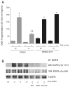
Fig.1
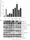
Fig.1. Effect of DFMO on apoptosis and EGFR activation
(A) Confluent serum starved cells were treated with 5 mM DFMO or DFMO (5mM) +putrescine (10 μM) for 1h followed by TNF-α/CHX for 3h. Cells were washed and DNA fragmentation was measured as described in the methods section. (mean ± SE, n=3, p<0.05considered significant). *, significantly different from TNF-α/CHX untreated, **, significantly different from TNF-α/CHX treated 0h. (B) Confluent serum starved cells were treated with 5 mM DFMO or DFMO (5mM) +putrescine (10μM), DFMO (5mM) + spermine (10μM), or DFMO+AG1478 for indicated time period and were washed and lysed using lysis buffer containing protease and phosphatase inhibitors. Whole cell lysates were subjected to immunoprecipitation using an EGFR specific antibody. The immunoprecipitates were washed 3 times with lysis buffer, subjected to SDS-PAGE and the membranes were probed with EGFR pY1173 and pY845 specific antibodies. The membranes were stripped and probed with EGFR specific antibody. Representative blots from 3 observations are shown.
Fig.2
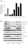
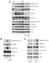
Fig.2. DFMO inhibits apoptosis by activating ERK1/2, integrin β3, Src and AKT
(A) Confluent serum starved cells were left untreated or pretreated with AG1478 (10 μM) or PP2 (10 μM) for 30 mins followed by DFMO (5 mM) or DFMO +putrescine (10μM) for 1h. These groups were then exposed to TNF-α/CHX for 3h. Cells were washed, and DNA fragmentation was measured as described in the methods section. (mean ± SE, n=3, p<0.05considered significant). *, significantly different from minus TNF-α/CHX UT, **, significantly different from TNF-α/CHX treated UT, †, significantly different from TNF-α/CHX treated DFMO group, ††, significantly different from TNF-α/CHX treated DFMO group or TNF-α/CHX treated UT group. (B) Confluent serum starved cells were treated with 5 mM DFMO for the indicated time period. A second group of cells pretreated with RGDS or AG1478 or PP2 were treated with DFMO for 5 min. A third group of cells were treated with DFMO (5mM) +putrescine (10μM) or DFMO (5mM) + spermine (10μM) for 5 min. Cells were washed and lysed using lysis buffer containing protease and phosphatase inhibitors. Whole cell lysates were subjected to SDS-PAGE and western blot analysis using phospho-specific ERK1/2, Src, AKT, and integrin β3 antibodies. The membranes were stripped and probed with respective antibodies recognizing total protein. The membranes were also stripped and probed with β-actin antibody. Representative blots from 3 observations are shown.
Fig.3
Fig.3. EGF protects cells against apoptosis via EGFR-mediated signaling
(A) Confluent serum starved cells left untreated (UT) or pretreated with AG1478 (10 μM) or PP2 (10 μM) for 30 mins were exposed to EGF (10 ng/ml) for additional 30 mins and incubated with TNF-α/CHX for 3h. Cells were washed and DNA fragmentation was measured as described in the methods section. (mean ± SE, n=3, p<0.05considered significant). *, significantly different from minus TNF-α/CHX UT, **, significantly different from TNF-α/CHX treated UT, †, significantly different from TNF-α/CHX treated EGF group, ††, significantly different from TNF-α/CHX treated EGF group and TNF-α/CHX treated UT group. (B) Confluent serum starved cells left untreated (UT) or pretreated with AG1478 (10 μM) or PP2 (10 μM) were exposed to EGF (10 ng/ml) for the indicated time period. Cells were washed and lysed using lysis buffer containing protease and phosphatase inhibitors. Whole cell lysates were subjected to SDS-PAGE and western blot analysis using phospho-specific ERK1/2, Src, and AKT antibodies. The membranes were stripped and probed with respective antibodies recognizing total protein. The membranes were also stripped and probed with an actin antibody. Representative blots from 3 observations are shown.
Fig.4
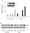
Fig.4. EGFR expression and apoptosis
(A) Cell lysates from NIH 3T3 EGFR knockout cells (PRN), PRN-cells expressing wild-type EGFR (WT), PRN-cells expressing K721A mutant EGFR (K721A), PRN-cells expressing Y845F mutant EGFR (Y845F) were subjected to SDS-PAGE and western blot analysis. The membrane was probed with EGFR antibody. The membrane was stripped and probed with β actin antibody. Representative blots from 3 observations are shown (Inset panel). NIH 3T3 EGFR knockout cells (PRN), PRN-cells expressing wild-type EGFR (WT), PRN-cells expressing K721A mutant EGFR (K721A), PRN-cells expressing Y845F mutant EGFR (Y845F) were grown in control and DFMO containing medium for 3 days and incubated for 24 h in the respective serum free medium. Cells were then exposed to TNF-α/CHX for 3h, washed, and DNA fragmentation was measured as described in the methods section. (mean ± SE, n=3, p<0.05considered significant). *, significantly different from TNF-α/CHX treated WT and PRN, †, significantly different from TNF-α/CHX treated control K721A. (B) The cell extracts from the above experiment were subjected to SDS-PAGE and western blot analysis. The membranes were probed with an antibody recognizing active caspase 3. The membranes were stripped and probed with β actin antibody. Representative blots from 3 observations are shown.
Fig.5
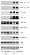
Fig.5. EGFR-mediated signaling in response to EGF and DFMO
Extracts prepared from confluent serum starved NIH 3T3 EGFR knockout cells (PRN) and PRN-cells expressing the wild-type EGFR (WT) cells left untreated or treated with EGF (10 ng/ml) or DFMO (5mM) for 3 mins were subjected to SDS-PAGE and western blot analysis. The membranes were probed with phospho specific EGFR, ERK1/2, Src, and AKT antibodies. The membranes were stripped and probed with antibodies recognizing respective total proteins and β actin. Representative blots from 3 observations are shown.
Fig.6
Fig.6. Fibronectin-induced Integrin β3 activation mediates signaling via the EGFR
IEC-6 cells seeded on plastic (PL) or fibronectin (FN)-coated plates were allowed to attach for the indicated time periods. Additional groups of cells plated on FN were exposed to putrescine (10μM) or spermine (5 μM) for 1h. Plates were washed with DPBS, and cells were lysed using lysis buffer containing protease and phosphatase inhibitors. The cell extracts were subjected to SDS-PAGE and western blot analysis. The membranes were probed with phospho specific EGFR, ERK1/2, integrin β3, Src, and AKT specific antibodies. The membranes were stripped and probed with antibodies recognizing respective total proteins and β actin (A). Representative blots from 3 observations are shown (A). IEC-6 (B), PRN, and WT (C) cells were allowed to attach on plastic or fibronectin-coated plates for 1h. One group of IEC-6 cells on FN plates was exposed to AG1478 (10μM) during attachment. Plates were washed with DPBS and cells were lysed using lysis buffer containing protease and phosphatase inhibitors. The cell extracts were subjected to SDS-PAGE and western blot analysis. The membranes were probed with phospho specific EGFR, ERK1/2, and Src, antibodies. The membranes were stripped and probed with antibodies recognizing respective total proteins and β actin. Representative blots from 3 observations are shown.
Fig.7
Fig.7. FN-mediated signaling protects cells against apoptosis
Confluent IEC-6 cells grown on plastic or FN-coated plates were left untreated or were treated with DFMO and exposed to TNF-α/CHX for 3h. Cells were washed, and DNA fragmentation was measured as described in the methods section (A). (mean ± SE, n=3, p<0.05considered significant). **, significantly different from TNF-α/CHX treated plastic group, †, significantly different from TNF-α/CHX untreated plastic.
Fig.8
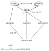
Fig.8. Schematic representation of polyamine-mediated signaling
Under basal conditions putrescine binds to the EGFR, integrin β3 and Src. Decreased putrescine during inhibition of ODC increases EGFR kinase and EGFR-mediated integrin β3 activities leading to activation of two major antiapoptotic signaling pathways MEK/ERK and Src. The stimulation of Src activates PI3K/AKT and JAK/STAT3 pathways. Together these pathways prevent TNF-α/CHX-induced apoptosis in IEC-6 cells.

Similar articles

Cited by

References

    1. Cheng H, Leblond CP. Am J Anat. 1974;141:537. - PubMed
    1. Schmidt GH, Winton DJ, Ponder BAJ. Development. 1988;103:785. - PubMed
    1. Potten CS, Loeffler MA. J Theor Biol. 1987;127:381. - PubMed
    1. Wright NA, Irwin M. Cell Tissue Kinet. 1982;15:595. - PubMed
    1. Grossman J, Walther K, Artinger M, Kiessling S, Schlomerich J. Cell Growth Differ. 2001;12:147. - PubMed

Publication types

MeSH terms

Substances