. 2007 Sep 18;17(18):1586-94.
doi: 10.1016/j.cub.2007.08.034. Epub 2007 Sep 6.

Disruption of intraflagellar transport in adult mice leads to obesity and slow-onset cystic kidney disease

Affiliations

Disruption of intraflagellar transport in adult mice leads to obesity and slow-onset cystic kidney disease

James R Davenport et al. Curr Biol. .

Abstract

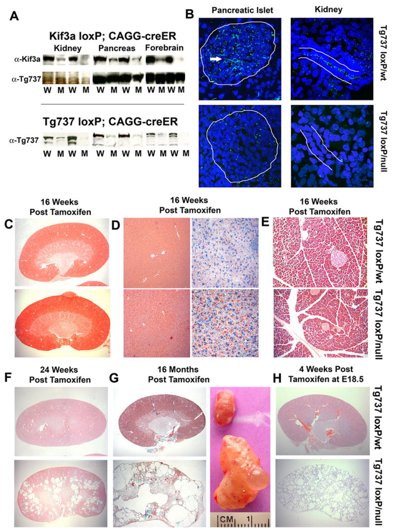
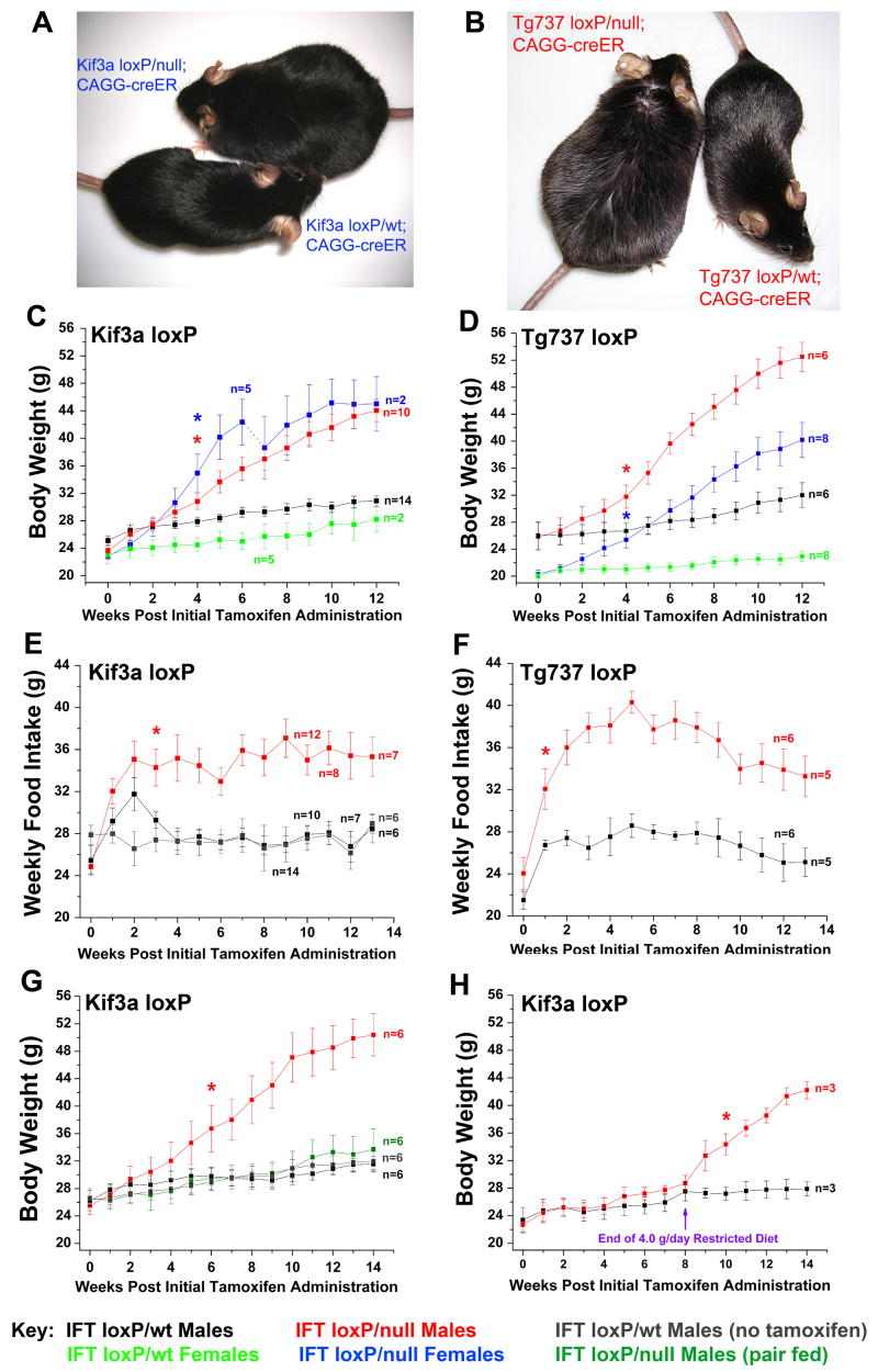
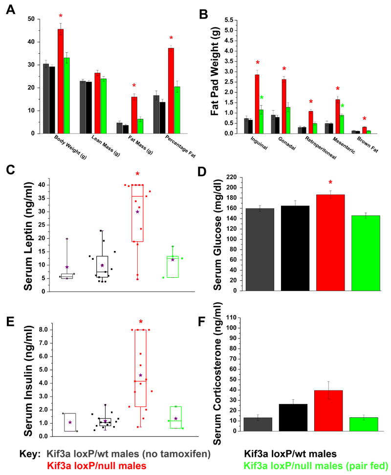
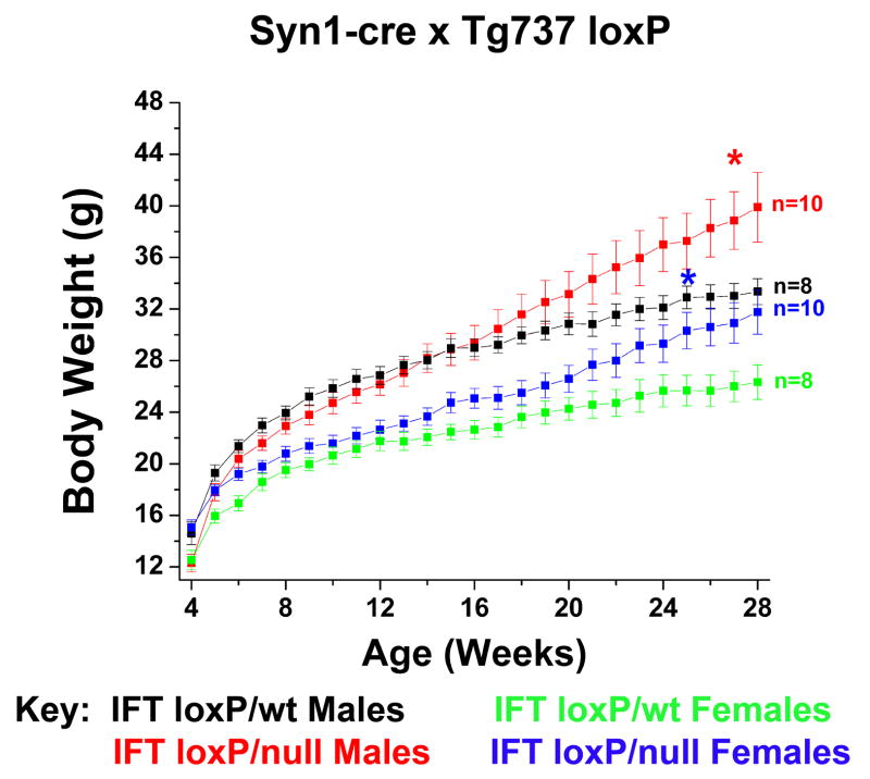
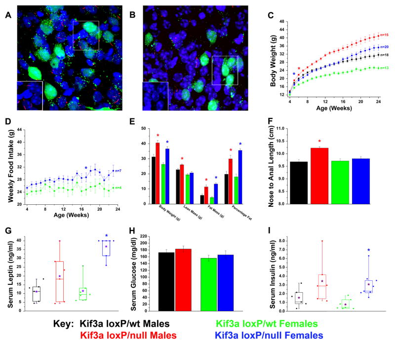
The assembly of primary cilia is dependent on intraflagellar transport (IFT), which mediates the bidirectional movement of proteins between the base and tip of the cilium. In mice, congenic mutations disrupting genes required for IFT (e.g., Tg737 or the IFT kinesin Kif3a) are embryonic lethal, whereas kidney-specific disruption of IFT results in severe, rapidly progressing cystic pathology. Although the function of primary cilia in most tissues is unknown, in the kidney they are mechanosenstive organelles that detect fluid flow through the tubule lumen. The loss of this flow-induced signaling pathway is thought to be a major contributing factor to cyst formation. Recent data also suggest that there is a connection between ciliary dysfunction and obesity as evidenced by the discovery that proteins associated with human obesity syndromes such as Alström and Bardet-Biedl localize to this organelle. To more directly assess the importance of cilia in postnatal life, we utilized conditional alleles of two ciliogenic genes (Tg737 and Kif3a) to systemically induce cilia loss in adults. Surprisingly, the cystic kidney pathology in these mutants is dependent on the time at which cilia loss was induced, suggesting that cyst formation is not simply caused by impaired mechanosensation. In addition to the cystic pathology, the conditional cilia mutant mice become obese, are hyperphagic, and have elevated levels of serum insulin, glucose, and leptin. We further defined where in the body cilia are required for normal energy homeostasis by disrupting cilia on neurons throughout the central nervous system and on pro-opiomelanocortin-expressing cells in the hypothalamus, both of which resulted in obesity. These data establish that neuronal cilia function in a pathway regulating satiety responses.


Figures

Figure 1
Figure 1. Systemic conditional deletion of Kif3a and Tg737 in adult mice leads to the development of slow onset cystic kidney disease
(A, top panel) Western blot analysis of Kif3a protein in kidney, pancreas, and brain in Kif3aloxP/wt; CAGG-creER™ (W) and Kif3aloxP/null; CAGG-creER™ (M) animals 16 weeks post tamoxifen administration. Tg737 protein expression (middle) was used as a loading control. (bottom panel) Tg737 protein expression was also measured in Tg737loxP; CAGG-creER™ mutant (M) and control (W) kidney, pancreas, and brain. (B) Confocal immunofluorescence analysis of Tg737-cWT and cKO tissues demonstrate loss of cilia in the pancreatic islet (left panels; 63x; Tg737-cWT above, Tg737-cKO below) and in the nephrons of the kidney (right; 100x; Tg737-cWT above, Tg737-cKO below). The islet and tubule are roughly outlined in white. The longer green staining pattern (pointed at by tip of white arrowhead) is due to background of the secondary antibody which reacts with an antigen on the vasculature and is unrelated to cilia. (C–E) Hematoxylin and Eosin (H&E) staining of (top) Tg737-cWT and (bottom) Tg737-cKO (C) kidney, (D, left panels) liver, and (E) pancreas at 16 weeks post tamoxifen administration in ad libitum fed mice shows no cystic pathology nor bile and pancreatic duct-related pathologies typical of the hypomorphic mutants at this age. Oil Red O staining (D right panels) shows an increase in lipid (red stain) accumulation in the mutants. (F) H&E staining of (top) Tg737-cWT and (bottom) Tg737-cKO kidneys at 24 weeks post tamoxifen injection in ad libitum fed mice revealed the development of cysts in Tg737-cKO mice (bottom). (G) Trichrome staining of (top) Tg737-cWT and (bottom) Tg737-cKO kidney sections 16 months post tamoxifen administration in ad libitum fed mice reveal the presence of very large cystic lesions throughout the kidney of the conditional mutants. Gross appearance of age and sex matched kidneys from Tg737-WT (top) and Tg737-cKO (bottom) are shown in the right panel. (H) H&E Staining of (top) Tg737-cWT and (bottom) Tg737-cKO kidney sections four weeks post administration of tamoxifen to the pregnant mother at embryonic day 18.5.
Figure 2
Figure 2. Systemic loss of Kif3a and/or Tg737 from adult mice results in a hyperphagia-induced obesity phenotype
(A) Images of Kif3a-cKO (left) and Kif3a-cWT (right) females 16 weeks after administration of tamoxifen. These littermates were of equivalent weight at time of injection. (B) Tg737-cKO (left) and Tg737-cWT (right) females are also shown 16 weeks after tamoxifen administration. (C,D) Body weight analysis of (C) Kif3a-cWT and Kif3a-cKO and (D) Tg737-cWT and Tg737-cKO males and females after tamoxifen administration (week zero). The number of Kif3a-cKO and Kif3a-cWT females analyzed in this study changed at week 6 when several of the mice were used to evaluate fat pad and organ weight masses. Animals were 8–12 weeks of age at the time of initial tamoxifen administration. (E,F) Weekly food intake analysis of (E) Kif3a-cKO and Kif3a-cWT males and (F) Tg737-cKO and Tg737-cWT males after initial tamoxifen administration (week 0). Several animals were euthanized in the course of the experiment for body composition and histological analyses. Kif3a-cWT controls that were not induced with tamoxifen are also shown in blue (E). (G) Body weight analysis in pair feeding studies of Kif3a-cKO males after administration of tamoxifen. Separate groups of Kif3a-cWT males were tested with or without tamoxifen to assess changes due to tamoxifen administration. (H) Kif3a-cWT and Kif3a-cKO males were administered tamoxifen and were diet restricted to 4.0 g/day for 8 subsequent weeks. At 8 weeks, all mice were then fed ad libitum (* = p≤0.05, indicates the initial point of significant deviation between controls and mutants).
Figure 3
Figure 3. Body composition and serum analysis of the obesity phenotype displayed in Kif3a-cKO males
(A) Dual energy X-ray absorptiometry (DXA) displaying lean mass, fat mass, and percentage body fat of male Kif3a-cWT and Kif3a-cKO mice 14 weeks after initial tamoxifen administration. Body weight was measured immediately after the DXA. (B) Carcass analysis of various fat pads and brown fat in Kif3a-cWT and Kif3a-cKO males 16 weeks after initial tamoxifen administration. (C) Box and whisker plot of non-fasting serum leptin levels in Kif3a-cWT and KO males 16 weeks after initial tamoxifen administration. 8 of 10 Kif3a-cKO ad libitum-fed mice were above the maximum threshold of detection (40 ng/ml) and were assigned a value of 40 ng/ml. Mean serum leptin levels are indicated by purple stars. (D) Serum glucose analysis of Kif3a-cWT and Kif3a-cKO males measured after 4 hours of fasting conditions. (E) Box and whisker plot of non-fasting serum insulin analysis of Kif3a-cWT and Kif3a-cKO males. 4 of 15 Kif3a-cKO males reached the highest detectable threshold for serum insulin analysis and were assigned the value of 8.0 ng/ml for statistical analysis. (F) Serum corticosterone analysis shows no significant differences between Kif3a-cWT and Kif3a-cKO males (* = p≤0.05, In (C) and (E), the means are indicated by the purple stars).
Figure 4
Figure 4. Conditional disruption of Tg737 in neurons
Body weights of Tg737-syn1KO and Tg737-syn1WT male and female mice indicate that loss of neuronal cilia due to disruption of Tg737 results in an obese phenotype.
Figure 5
Figure 5. Conditional deletion of Kif3a from POMC-expressing cells in mice leads to increases in weight, adiposity, and body length
(A, B) Confocal immunofluorescence analysis of (A) Kif3aloxP/wt (Kif3a-pomcWT) and (B) Kif3aloxP/null (Kif3a-pomcKO) arcuate nucleus (100x) bred onto the Z/EG Cre reporter line in the presence of the POMC-cre deletor strain demonstrates the loss or stunting of neuronal cilia (red) on affected POMC neurons (green). Inserts show cilia (red) without the GFP signal and are derived from the boxed region in the image. Note that cilia are still retained on the non-POMC expressing cells in the hypothalamus. (C) Body weight analysis of Kif3a-pomcKO and Kif3a-pomcWT males and females. (D) Weekly food intake of Kif3a-pomcKO females was consistently increased from their Kif3a-pomcWT controls. (E) DXA analysis performed on adult (age 22–25 weeks) mice showed significant increases in fat mass and percentage fat in both sexes, and lean mass in male Kif3a-pomcKO mice. Body weight was measured after the DXA analysis. (F) Nose-to-anal length analysis conducted in anesthetized adult (age 22–25 weeks) Kif3a-pomcWT and Kif3a-pomcKO mice. The average length from independently determined two blinded measurements was used. (G–I) Serum analysis of (G) non-fasted leptin, (H) four-hour fasted glucose, and (I) non-fasted insulin in Kif3a-pomcWT and Kif3a-pomcKO mice (* = p≤0.05, in (C) and (D) indicating the initial point of significant deviation between controls and mutants. The purple stars in G and I represent the means of the individual groups.

Similar articles

Cited by

References

    1. Lin F, Hiesberger T, Cordes K, Sinclair AM, Goldstein LS, Somlo S, Igarashi P. Kidney-specific inactivation of the KIF3A subunit of kinesin-II inhibits renal ciliogenesis and produces polycystic kidney disease. Proc Natl Acad Sci U S A. 2003;100:5286–5291. - PMC - PubMed
    1. Marszalek JR, Ruiz-Lozano P, Roberts E, Chien KR, Goldstein LS. Situs inversus and embryonic ciliary morphogenesis defects in mouse mutants lacking the KIF3A subunit of kinesin-II. Proc Natl Acad Sci U S A. 1999;96:5043–5048. - PMC - PubMed
    1. Murcia NS, Richards WG, Yoder BK, Mucenski ML, Dunlap JR, Woychik RP. The Oak Ridge Polycystic Kidney (orpk) disease gene is required for left-right axis determination. Development. 2000;127:2347–2355. - PubMed
    1. Singla V, Reiter JF. The primary cilium as the cell's antenna: signaling at a sensory organelle. Science. 2006;313:629–633. - PubMed
    1. Praetorius HA, Spring KR. The renal cell primary cilium functions as a flow sensor. Curr Opin Nephrol Hypertens. 2003;12:517–520. - PubMed

Publication types

MeSH terms