

Evidence for acquired pregenual anterior cingulate gray matter loss from a twin study of combat-related posttraumatic stress disorder
- PMID: 17825801
- PMCID: PMC2752671
- DOI: 10.1016/j.biopsych.2007.06.022
Evidence for acquired pregenual anterior cingulate gray matter loss from a twin study of combat-related posttraumatic stress disorder
Abstract
Background: Controversy exists over the nature and origin of reduced regional brain volumes in posttraumatic stress disorder (PTSD). At issue is whether these reductions represent preexisting vulnerability factors for developing PTSD upon traumatic exposure or acquired PTSD signs due to the traumatic stress that caused the PTSD or the chronic stress of having the disorder (or both). We employed a case-control design in monozygotic twin pairs discordant for combat exposure to address the preexisting versus acquired origin of brain morphometric abnormalities in PTSD.
Methods: We used voxel-based morphometry to search for gray matter density reductions in magnetic resonance imaging (MRI) data obtained in a previous study of combat-exposed Vietnam veteran twins with (n = 18) versus without (n = 23) PTSD and their "high-risk" versus "low-risk" (respectively) identical combat-unexposed cotwins.
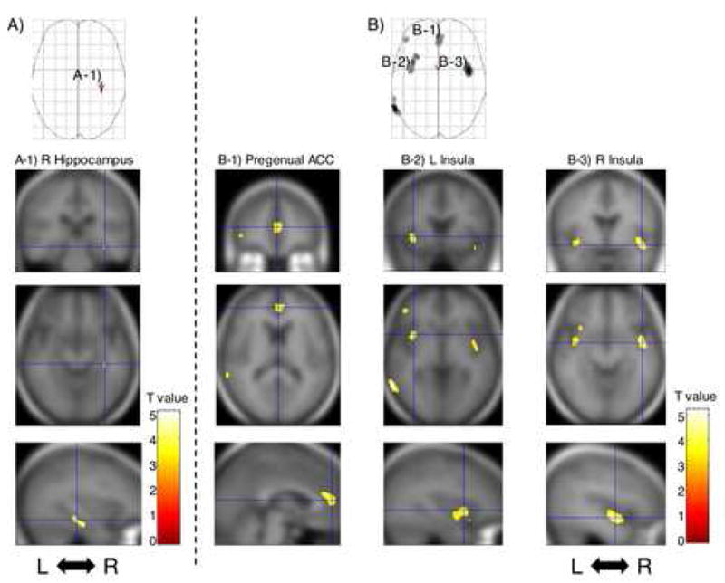
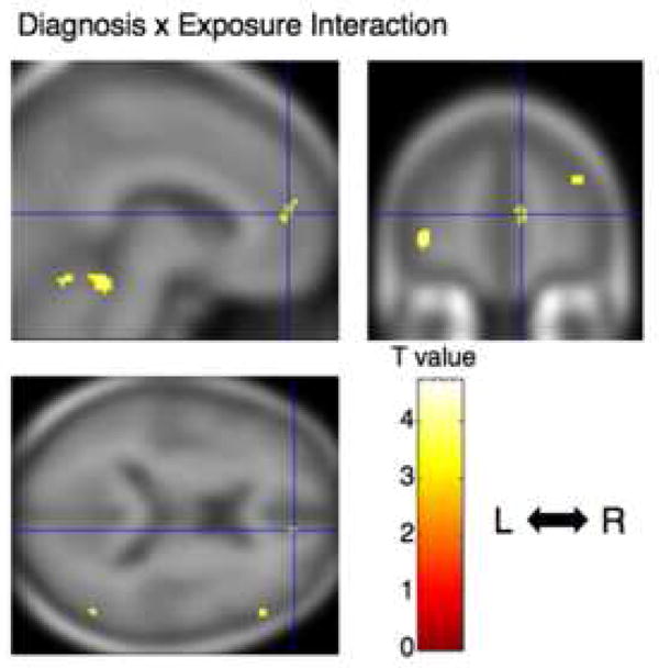
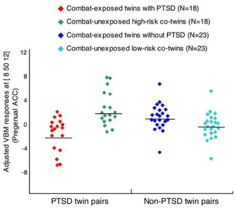
Results: Compared with the combat-exposed twins without PTSD, the combat-exposed twins with PTSD showed significant gray matter density reductions in four predicted brain regions: right hippocampus, pregenual anterior cingulate cortex (ACC), and left and right insulae. There was a significant PTSD Diagnosis x Combat Exposure interaction in pregenual ACC in which combat-exposed PTSD twins had lower gray matter density than their own combat-unexposed cotwins as well as than the combat-exposed twins without PTSD and their cotwins.
Conclusions: The results point to gray matter volume diminutions in limbic and paralimbic structures in PTSD. The pattern of results obtained for pregenual ACC suggests that gray matter reduction in this region represents an acquired sign of PTSD consistent with stress-induced loss.
Figures
Comment in
-
Regional specificity of traumatic stress-related cortical reduction: further evidence from a twin study of post-traumatic stress disorder.Biol Psychiatry. 2008 Mar 15;63(6):539-41. doi: 10.1016/j.biopsych.2008.01.015. Biol Psychiatry. 2008. PMID: 18295657 No abstract available.
Similar articles
-
Exaggerated activation of dorsal anterior cingulate cortex during cognitive interference: a monozygotic twin study of posttraumatic stress disorder.Am J Psychiatry. 2011 Sep;168(9):979-85. doi: 10.1176/appi.ajp.2011.09121812. Epub 2011 Jul 1. Am J Psychiatry. 2011. PMID: 21724666 Free PMC article.
-
Regional specificity of traumatic stress-related cortical reduction: further evidence from a twin study of post-traumatic stress disorder.Biol Psychiatry. 2008 Mar 15;63(6):539-41. doi: 10.1016/j.biopsych.2008.01.015. Biol Psychiatry. 2008. PMID: 18295657 No abstract available.
-
Subtle neurologic compromise as a vulnerability factor for combat-related posttraumatic stress disorder: results of a twin study.Arch Gen Psychiatry. 2006 May;63(5):571-6. doi: 10.1001/archpsyc.63.5.571. Arch Gen Psychiatry. 2006. PMID: 16651514
-
Clarifying the origin of biological abnormalities in PTSD through the study of identical twins discordant for combat exposure.Ann N Y Acad Sci. 2006 Jul;1071:242-54. doi: 10.1196/annals.1364.019. Ann N Y Acad Sci. 2006. PMID: 16891575 Free PMC article.
-
Twin studies of posttraumatic stress disorder: differentiating vulnerability factors from sequelae.Neuropharmacology. 2012 Feb;62(2):647-53. doi: 10.1016/j.neuropharm.2011.03.012. Epub 2011 Mar 31. Neuropharmacology. 2012. PMID: 21443892 Free PMC article. Review.
Cited by
-
Voxel-based morphometric gray matter correlates of posttraumatic stress disorder.J Anxiety Disord. 2013 May;27(4):413-9. doi: 10.1016/j.janxdis.2013.04.004. Epub 2013 Apr 26. J Anxiety Disord. 2013. PMID: 23746489 Free PMC article.
-
Trauma-specific Grey Matter Alterations in PTSD.Sci Rep. 2016 Sep 21;6:33748. doi: 10.1038/srep33748. Sci Rep. 2016. PMID: 27651030 Free PMC article. Review.
-
Vulnerability of the Hippocampus to Insults: Links to Blood-Brain Barrier Dysfunction.Int J Mol Sci. 2024 Feb 6;25(4):1991. doi: 10.3390/ijms25041991. Int J Mol Sci. 2024. PMID: 38396670 Free PMC article. Review.
-
Altered processing of contextual information during fear extinction in PTSD: an fMRI study.CNS Neurosci Ther. 2011 Aug;17(4):227-36. doi: 10.1111/j.1755-5949.2010.00152.x. Epub 2010 Apr 16. CNS Neurosci Ther. 2011. PMID: 20406268 Free PMC article.
-
Risk of diabetes in U.S. military service members in relation to combat deployment and mental health.Diabetes Care. 2010 Aug;33(8):1771-7. doi: 10.2337/dc10-0296. Epub 2010 May 18. Diabetes Care. 2010. PMID: 20484134 Free PMC article.
References
-
- Kitayama N, Vaccarino V, Kutner M, Weiss P, Bremner JD. Magnetic resonance imaging (MRI) measurement of hippocampal volume in posttraumatic stress disorder: a meta-analysis. J Affect Disord. 2005;88:79–86. - PubMed
-
- Rauch SL, Shin LM, Segal E, Pitman RK, Carson MA, McMullin K, Whalen PJ, Makris N. Selectively reduced regional cortical volumes in post-traumatic stress disorder. Neuroreport. 2003;14:913–916. - PubMed
-
- Milad MR, Rauch SL, Pitman RK, Quirk GJ. Fear extinction in rats: implications for human brain imaging and anxiety disorders. Biol Psychol. 2006;73:61–71. - PubMed
Publication types
MeSH terms
Grants and funding
LinkOut - more resources
Full Text Sources
Medical
