

Tissue kallikrein and kinin infusion rescues failing myocardium after myocardial infarction
- PMID: 17826650
- PMCID: PMC4519013
- DOI: 10.1016/j.cardfail.2007.04.009
Tissue kallikrein and kinin infusion rescues failing myocardium after myocardial infarction
Abstract
Background: Tissue kallikrein is a serine proteinase that generates the vasoactive kinin peptide, which produces vasodilatory, angiogenic, and antiapoptotic effects. In this study, we investigated the effect of a stable supply of kallikrein and kinin on ventricular remodeling and blood vessel growth in rats after myocardial infarction.
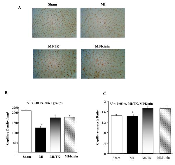
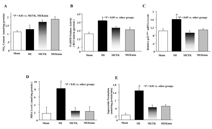
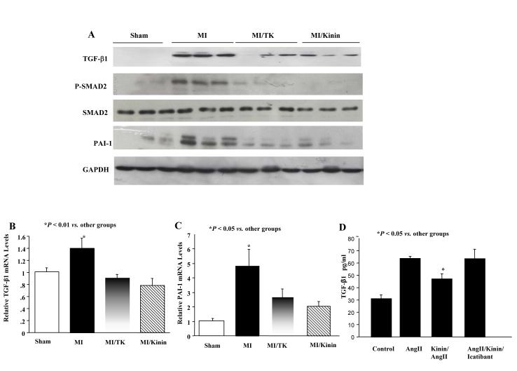
Methods and results: At 1 week after coronary artery ligation, tissue kallikrein or kinin was infused through a minipump for 4 weeks. At 5 weeks after myocardial infarction, kallikrein and kinin infusion significantly improved cardiac contractility and reduced diastolic dysfunction without affecting systolic blood pressure. Kallikrein and kinin infusion significantly increased capillary density in the noninfarcted region. Kallikrein and kinin infusion also reduced heart weight/body weight ratio, cardiomyocyte size, and atrial natriuretic peptide and brain natriuretic peptide expression in the noninfarcted area. Moreover, kallikrein and kinin infusion inhibited interstitial collagen deposition, collagen fraction volume, and collagen I and collagen III mRNA levels, transforming growth factor (TGF)-beta1 and plasminogen activator inhibitor-1 expression, and Smad2 phosphorylation. The effects of kallikrein and kinin on cardiac remodeling were associated with increased nitric oxide levels and reduced NADPH oxidase expression and activity, superoxide formation, and malondialdehyde levels. Furthermore, in cultured cardiac fibroblasts, kinin inhibited angiotensin II-stimulated TGF-beta1 production, and the effect was blocked by icatibant.
Conclusion: These results indicate that a subdepressor dose of kallikrein or kinin can restore impaired cardiac function in rats with postinfarction heart failure by inhibiting hypertrophy and fibrosis and promoting angiogenesis through increased nitric oxide formation and suppression of oxidative stress and TGF-beta1 expression.
Figures


References
-
- Paul M, Stock P, Langheinrich M, Liefeldt L, Schonfelder G, Bohm M. Role of the cardiac renin-angiotensin system in human heart failure. Adv Exp Med Biol. 1995;377:279–83. - PubMed
-
- Hill MF, Singal PK. Right and left myocardial antioxidant responses during heart failure subsequent to myocardial infarction. Circulation. 1997;96:2414–20. - PubMed
-
- Lu L, Quinn MT, Sun Y. Oxidative stress in the infarcted heart: role of de novo angiotensin II production. Biochem Biophys Res Commun. 2004;17:943–51. - PubMed
-
- Hogg N, Browning J, Howard T, Winterford C, Fitzpatrick D, Gobe G. Apoptosis in vascular endothelial cells caused by serum deprivation, oxidative stress and transforming growth factor-beta. Endothelium. 1999;7:35–49. - PubMed
MeSH terms
Substances
Grants and funding
LinkOut - more resources
Full Text Sources
Medical
