. 2007 Oct 15;310(2):317-28.
doi: 10.1016/j.ydbio.2007.07.042. Epub 2007 Aug 15.

Embryonic origin of gustatory cranial sensory neurons

Affiliations

Embryonic origin of gustatory cranial sensory neurons

Danielle E Harlow et al. Dev Biol. .

Abstract

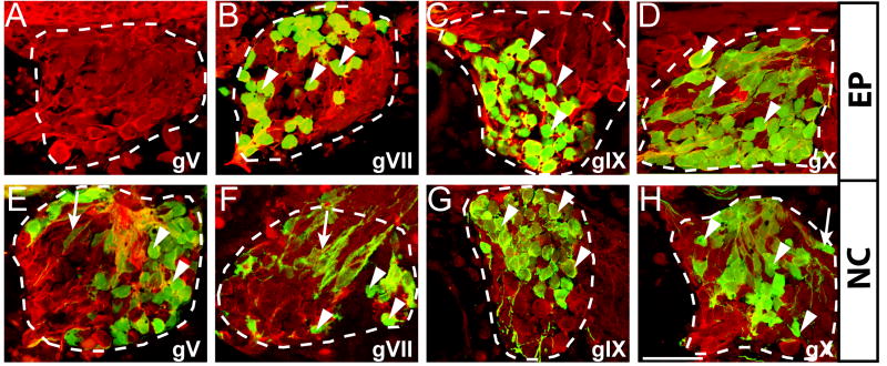
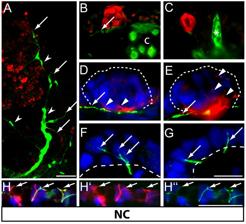
Cranial nerves VII, IX and X provide both gustatory (taste) and non-gustatory (touch, pain, temperature) innervation to the oral cavity of vertebrates. Gustatory neurons innervate taste buds and project centrally to the rostral nucleus of the solitary tract (NTS), whereas neurons providing general epithelial innervation to the oropharynx project to non-gustatory hindbrain regions, i.e., spinal trigeminal nucleus. In addition to this dichotomy in function, cranial ganglia VII, IX and X have dual embryonic origins, comprising sensory neurons derived from both cranial neural crest and epibranchial placodes. We used a fate mapping approach to test the hypothesis that epibranchial placodes give rise to gustatory neurons, whereas the neural crest generates non-gustatory cells. Placodal ectoderm or neural crest was grafted from Green Fluorescent Protein (GFP) expressing salamander embryos into unlabeled hosts, allowing us to discern the postembryonic central and peripheral projections of each embryonic neuronal population. Neurites that innervate taste buds are exclusively placodal in origin, and their central processes project to the NTS, consistent with a gustatory fate. In contrast, neural crest-derived neurons do not innervate taste buds; instead, neurites of these sensory neurons terminate as free nerve endings within the oral epithelium. Further, the majority of centrally directed fibers of neural crest neurons terminate outside the NTS, in regions that receive general epithelial afferents. Our data provide empirical evidence that embryonic origin dictates mature neuron function within cranial sensory ganglia: specifically, gustatory neurons derive from epibranchial placodes, whereas neural crest-derived neurons provide general epithelial innervation to the oral cavity.


Figures

Figure 1
Figure 1. Experimental approach for fate mapping of epibranchial placode and neural crest cells
(A) Grafts were taken from GFP mRNA microinjected or transgenic GFP-expressing donor embryos. (B) Schematic diagram of transplantation of epibranchial placodal ectoderm (EP) from stage 19 GFP-labeled donor into unlabeled host embryo. (C) Diagram of GFP-labeled neural crest (NC) transplantation in stage 17 embryos. A small piece of the dorsal neural fold containing premigratory neural crest cells of the cranial ganglia (area between dashed white line) as well as cells destined to give rise to dorsal hindbrain were transplanted. (D) GFP-labeled placodal ectoderm or (F) neural fold graft recipients one-hour post-surgery. The embryo and the dorsal neural fold are outlined in white. Orientation is identical to B&C. (E) In an ectoderm graft recipient, 8 days later (stage 41), GFP-labeled nerves (arrows) in the jaw are visible in lateral view. The eye (“e”) is autofluorescent and is not labeled with GFP. (G) In a stage 41 neural fold graft recipient, GFP-labeled cartilage and muscle (asterisks) as well as GFP-labeled nerves (arrows) are evident in the jaw. In all images, anterior is right, and dorsal is up. GFP = green. Scale bars: 2mm for A-D, F; 500 μm for E and G.
Figure 2
Figure 2. Epibranchial placodes contribute neurons to ganglia VII, IX, and X, while the neural crest contributes both neurons and glia
Confocal z-stacks through sagittal sections of ganglia V (gV), VII (gVII), IX (gIX) and X (gX) reveal the presence of GFP-labeled cells. (A-D) In sections from larvae with epibranchial placode grafts, GFP labels neurons (arrowheads) within gVII (B), gVII (C) and gX (D), but does not label neurons in ganglion V. (E-H) In larvae with NC grafts, neural crest derivatives within the ganglia include both neurons (arrowheads) and glial cells (arrows) in gV (E), gVII (F), gIX (G) and gX (H). Labeled neurons (arrowheads) have characteristic large round nuclei and are immunoreactive for acetylated tubulin and GFP. GFP-labeled satellite glial cells (arrows) can be seen ensheathing sensory neurons. Ganglia are outlined with a dashed line. Dorsal is up, anterior is to the left. Anti-GFP = green; anti-acetylated tubulin = red. Scale bar = 50 μm.
Figure 3
Figure 3. Sensory neurons derived from epibranchial placodes innervate taste buds
(A) Confocal z-stack of the interior of the upper jaw of an epibranchial placode GFP-graft recipient at stage 41. GFP-positive rami of the VIIth nerve (arrows) branch to contact taste buds labeled with anti-calretinin (red). Anterior is to the top. (B-D) Transverse sections through calretinin-positive taste buds reveal that GFP-labeled fibers (green) form a large nerve plexus beneath each taste bud and extend fine processes (arrows) apically into the buds. (E) GFP-positive fibers (arrows) within taste buds (outlined by white dashed lines) are also positive for anti-acetylated tubulin (AT, red) in (F). (G) Merged image shows that GFP label co-localizes with AT. Nuclei are stained blue with Hoechst. Anti-GFP = green. Scale bar in A = 100μm; B-G = 25 μm.
Figure 4
Figure 4. Sensory neurons derived from neural crest innervate non-taste bud-bearing epithelium in the oral cavity
(A) Confocal z-stack of the interior of upper jaw of neural crest GFP-graft recipient at stage 41, reveals GFP-positive nerve fibers (arrows) terminating in oral epithelium devoid of taste buds. GFP-positive Schwann cells (arrowheads) ensheath these peripheral nerves. (B-C) In transverse sections, several expected neural crest derivatives are labeled with GFP, including nerve fibers (arrow in B), cartilage (‘c’ in B) and odontoblasts (asterisk in C). No GFP-labeled nerve fibers were present in calretinin (red) positive taste buds. (D-E) GFP-labeled crest-derived nerve fibers (arrows) travel beneath taste buds (outlined with dashed line), but do not project into taste buds. AT-positive fibers (red) within taste buds (arrowheads) are not labeled with GFP. (F-G). Instead, GFP-positive fibers of neural crest neurons terminate as free nerve endings (arrows in F, G) within the non-taste-bud-bearing oropharyngeal epithelium. Dashed line indicates basement membrane. (H) Merged image shows that some of the AT-positive (red, H‘) nerve fibers in the skin above the otic vesicle are also neural crest derived as evident by double-labeling with GFP (green, H“). Anti-GFP=green. Nuclei are stained blue with Hoechst in D-H. Scale bar = 100 μm for A; 50 for B-C, and 25 μm for D-H.
Figure 5
Figure 5. Placode-derived sensory neurons project to the solitary tract nucleus via the fasciculus solitarius, while neural crest-derived sensory neurons do not
Top panel is a diagram of a lateral view of a larval axolotl brain, anterior to the right, with lines indicating the level of the transverse sections shown in A-F below. (A-C) Central projections of GFP-labeled placodal neurons (green) are only seen in the fasciculus solitarius (f.sol.) shown here at the level of the (A) VIIth, (B) IXth, and (C) Xth nerve roots on the operated side of placodal ectoderm larval graft recipients. (D-F) Central projections of GFP-labeled neural crest neurons were not present in (D) the fasciculus solitarius rostrally at the level of the VIIth root or more caudally at the level of the (E) IXth or (F) Xth roots, but rather were present in other specific hindbrain regions, including the spinal trigeminal tract (SpV). Central afferents of coincidentaly labeled lateral line neurons (r.l.l.) were sometimes seen in both placode (C) and crest (F) graft recipeints. Anterior is up. Nuclei are stained blue with Hoechst. Anti-GFP = green. f.sol. = fasciulus solitarius; r.l.l. = roots of the lateral line nerves; r.IX & rX = roots of IX and X; SpV = spinal trigeminal tract. Scale bar=20μm.

References

    1. Adelmann HB. The development of the neural folds and cranial ganglia of the rat. Journal of Comparative Neurology. 1925;39:19–171.
    1. Agerman K, Hjerling-Leffler J, Blanchard MP, Scarfone E, Canlon B, Nosrat C, Ernfors P. BDNF gene replacement reveals multiple mechanisms for establishing neurotrophin specificity during sensory nervous system development. Development. 2003;130:1479–91. - PubMed
    1. Altman JS, Dawes EA. A cobalt study of medullary sensory projections from lateral line nerves, associated cutaneous nerves, and the VIIIth nerve in adult Xenopus. J Comp Neurol. 1983;213:310–26. - PubMed
    1. Artinger KB, Fedtsova N, Rhee JM, Bronner-Fraser M, Turner E. Placodal origin of Brn-3-expressing cranial sensory neurons. Journal of Neurobiology. 1998;36:572–85. - PubMed
    1. Arvidsson J, Fundin BT, Pfaller K. Innervation of the hard palate in the rat studied by anterograde transport of horseradish peroxidase conjugates. The Journal of Comparative Neurology. 1995;351:489–498. - PubMed

Publication types