. 2007 Oct 15;310(2):379-87.
doi: 10.1016/j.ydbio.2007.08.021. Epub 2007 Aug 16.

Cessation of renal morphogenesis in mice

Affiliations

Cessation of renal morphogenesis in mice

Heather A Hartman et al. Dev Biol. .

Abstract

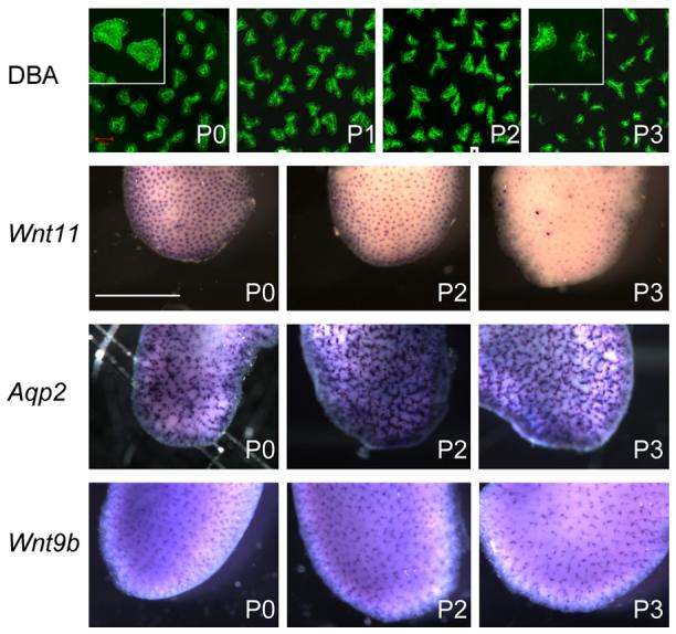
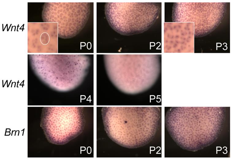
The kidney develops by cycles of ureteric bud branching and nephron formation. The cycles begin and are sustained by reciprocal inductive interactions and feedback between ureteric bud tips and the surrounding mesenchyme. Understanding how the cycles end is important because it controls nephron number. During the period when nephrogenesis ends in mice, we examined the morphology, gene expression, and function of the domains that control branching and nephrogenesis. We found that the nephrogenic mesenchyme, which is required for continued branching, was gone by the third postnatal day. This was associated with an accelerated rate of new nephron formation in the absence of apoptosis. At the same time, the tips of the ureteric bud branches lost the typical appearance of an ampulla and lost Wnt11 expression, consistent with the absence of the capping mesenchyme. Surprisingly, expression of Wnt9b, a gene necessary for mesenchyme induction, continued. We then tested the postnatal day three bud branch tip and showed that it maintained its ability both to promote survival of metanephric mesenchyme and to induce nephrogenesis in culture. These results suggest that the sequence of events leading to disruption of the cycle of branching morphogenesis and nephrogenesis began with the loss of mesenchyme that resulted from its conversion into nephrons.


Figures

Fig.1
Fig.1. Capping nephrogenic mesenchyme rapidly converted to nephrons in the immediate postnatal period
Whole mount immunostaining with antibody to CITED1 and imaging by confocal microscopy demonstrates large clusters of nephrogenic mesenchyme (red) at birth (P0). The patches of nephrogenic mesenchyme were smaller by P1 and were scarce by P2. (The images are merged images of all Z-stack slices from the surface of the kidney to a depth of 35μ.) In situ hybridization for Six2 demonstrated the same change in capping mesenchyme. There were no apparent changes in patterns of proliferation or apoptosis. Staining with antibody to PHH3 (red) identified proliferating cells within the mesenchyme, nephrons, and ureeric bud at E16.5 and P0. Because the capping mesenchyme was nearly absent by P2, the proliferation that was seen was mostly in developing nephrons rather than in the mesenchyme. Proliferation also continued in the ureteric bud. E-cadherin expression (blue) was used to help identify structures and was seen within ureteric bud derivatives and more mature nephrons. (Images are single slices at a depth of 25μ for E16.5 and P0, and at a depth of 16μ for P2 and P3). Staining of P0 kidneys with antibody to cleaved Caspase 3 (red) showed only sparse apoptotic activity in the surface nephrogenic region (0-25μ) in mesenchymal or ureteric bud cells at all ages examined. In contrast, apoptotic activity was common deeper within the kidney (30-55μ). (Images are merged images of all slices of a Z-stack series between the surface and 25μ and between 30 and 55μ. The magnification of all confocal microscopy images is the same. green bar = 50μ) Whole mount in situ hybridization showed diminished expression of Foxd1 in the postnatal day 3 kidney. During the first 3 postnatal days, WT-1 stained renal vesicles replace the metanephric mesenchyme that had surrounded the ureteric bud branch tips. The staining pattern of the mesenchyme with antibody to WT1 is similar in the embryonic (E16.5) and P0 kidney. By P2 renal vesicles (arrows) replace the nephrogenic mesenchyme near the surface of the kidney. By P3 those vesicles develop further and show polarized expression of WT1 (arrowhead) opposite the ureteric bud tip. (Circles were placed around vesicles or glomerular anlagen to illustrate the manner in which these structures were measured. The images are single optical slices at a depth of 20μ from the surface for E16.5 and 15μ for the rest.) A similar pattern was seen for Pax2 which is expressed in the ureteric bud, capping mesenchyme and early nephron. The mesenchyme staining remained similar in intensity to that in the tips and early nephrons, and demonstrated the replacement of the mesenchyme around the tips by nascent nephrons (15μ from the surface).
Fig. 2
Fig. 2. Ureteric bud branch tip morphology and gene expression pattern changed in the early postnatal period
The morphology of the ureteric bud branch tips, stained with DBA (green), changed from larger rounded tips to smaller scalloped tips in the postnatal period (insets - higher magnification). The tips still remain near the surface of the kidney. (The images are merged images of Z-stack slices from the surface of DBA stained kidneys to a depth of 35μ. All images are the same magnification. red bar = 50μ) In situ hybridization showed that the expression of Wnt11 in the ureteric bud branch tips is down-regulated in the postnatal period whereas the expression of Wnt9b and Aqp2, two other markers in ureteric bud branches, remained constant. White bar=1mm
Fig. 3
Fig. 3. The postnatal day 3 ureteric bud branch tip still had the potential to induce nephrons and promote survival of metanephric mesenchyme
A. Ureteric bud branch tips, which express E-cadherin (blue), induced nephrons from GFP-positive mesenchyme. The more mature tubules of these in vitro-induced nephrons expressed both GFP (green) and E-cadherin (blue), while the glomerular anlage expressed both WT1 (red) and GFP (green). The glomerular anlage can be identified by the characteristic hemispheric shape and double contour of cells. The arrows indicate the junction where the GFP-positive nephron connects with the ureteric bud. bar= 50μ B. After 4 days in culture, a rim of E11.5-derived metanephric mesenchyme (arrow), which expresses GFP (green) and WT1 (red), surrounds a ureteric bud branch tip (blue) obtained from a P3 kidney. Note the weak GFP signal in the ureteric bud (arrowhead). Bar=100μ
Fig. 4
Fig. 4. Nephron density increased and mature nephrons were located more superficially in the postnatal period
A. Nephron density continues to increase in the postnatal period (*p< .001 compared to E16.5 and P2, t-test, n=5). The increase in density was greater in the 2 days between birth and P2 (202 nephrons/mm2 increase) than it was in the 3 days between E16.5 and birth (153 nephrons/mm2 increase). The density appeared to reach a plateau by P3. B. The relationship of nephron maturity, as determined by vesicle or glomerular anlage size, to their depth within the kidney was similar at E16.5 and P0. Afterward, more mature nephrons were found nearer the surface of the kidney. The correlation between glomerular anlage size and depth decreased resulting in a change in the R2 value from .66 on P0 to .21 on P2 (n=5). In these figures, the diameter of the WT1 stained portion of the nascent nephron was plotted against the depth from the surface.
Fig. 5
Fig. 5. The increased density of newly induced nephrons can be seen using a marker for pretubular aggregates
Wnt4 expression in pretubular aggregates appears restricted in patches around the bud tips on P0, is spread more evenly by P2, and is extinguished by P5. A cluster of aggregates is circled in the inset of P0 kidney. Similar clusters cannot be identified on P3. The expression pattern contrasts what is seen using a marker for more mature nephron structures. The pattern of Brn-1, a marker of distal tubules, changed little in the immediate postnatal period.

References

    1. Carroll TJ, et al. Wnt9b plays a central role in the regulation of mesenchymal to epithelial transitions underlying organogenesis of the mammalian urogenital system. Dev Cell. 2005;9:283–92. - PubMed
    1. Chuang PT, McMahon AP. Branching morphogenesis of the lung: new molecular insights into an old problem. Trends Cell Biol. 2003;13:86–91. - PubMed
    1. Dudley AT, et al. Interaction between FGF and BMP signaling pathways regulates development of metanephric mesenchyme. Genes Dev. 1999;13:1601–13. - PMC - PubMed
    1. Durbec P, et al. GDNF signalling through the Ret receptor tyrosine kinase. Nature. 1996;381:789–93. - PubMed
    1. Grieshammer U, et al. FGF8 is required for cell survival at distinct stages of nephrogenesis and for regulation of gene expression in nascent nephrons. Development. 2005;132:3847–57. - PubMed

Publication types

LinkOut - more resources