. 2007 Sep-Nov;29(2-3):174-86.
doi: 10.1016/j.jaut.2007.07.005.

Species-specific immune responses generated by histidyl-tRNA synthetase immunization are associated with muscle and lung inflammation

Affiliations

Species-specific immune responses generated by histidyl-tRNA synthetase immunization are associated with muscle and lung inflammation

Yasuhiro Katsumata et al. J Autoimmun. 2007 Sep-Nov.

Abstract

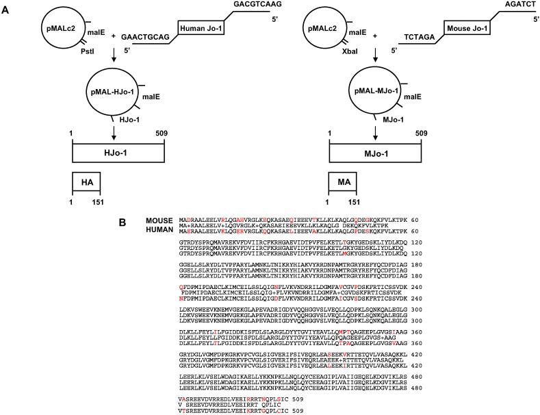
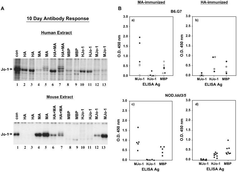
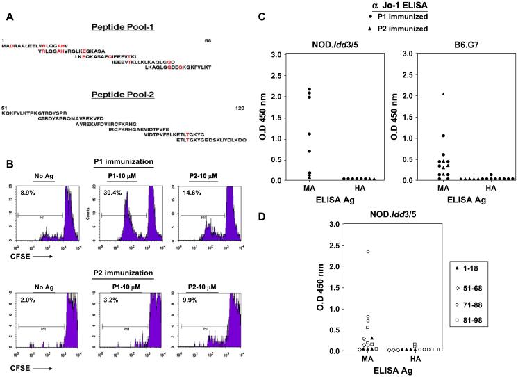
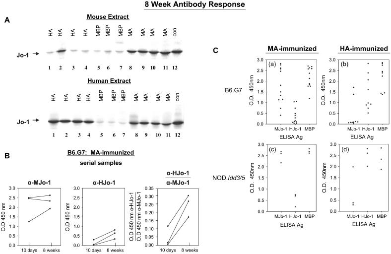
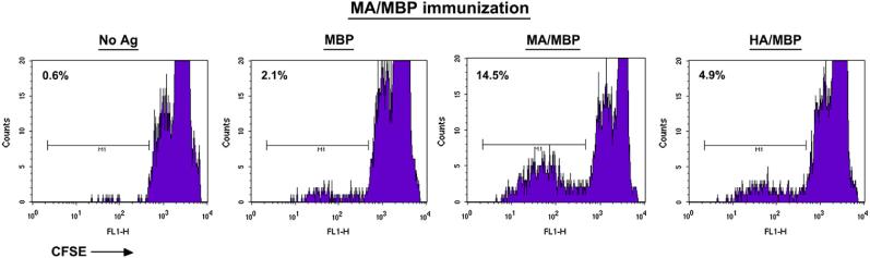
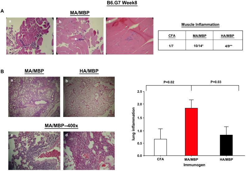
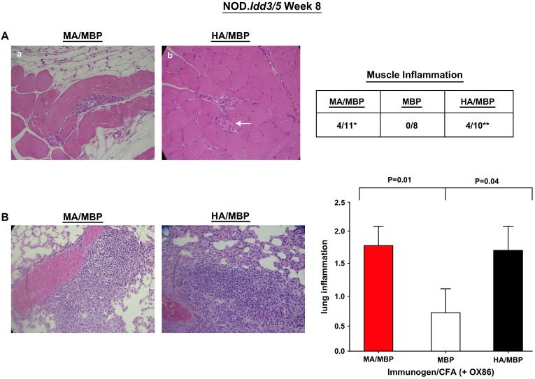
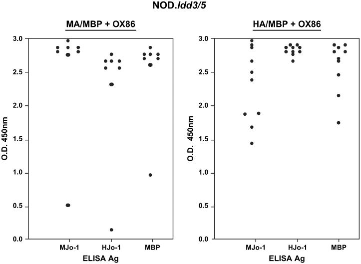
Evidence implicating histidyl-tRNA synthetase (Jo-1) in the pathogenesis of the anti-synthetase syndrome includes established genetic associations linking the reproducible phenotype of muscle inflammation and interstitial lung disease with autoantibodies recognizing Jo-1. To better address the role of Jo-1-directed B and T cell responses in the context of different genetic backgrounds, we employed Jo-1 protein immunization of C57BL/6 and NOD congenic mice. Detailed analysis of early antibody responses following inoculation with human or murine Jo-1 demonstrates remarkable species-specifity, with limited cross recognition of Jo-1 from the opposite species. Complementing these results, immunization with purified peptides derived from murine Jo-1 generates B and T cells targeting species-specific epitopes contained within the amino terminal 120 amino acids of murine Jo-1. The eventual spreading of B cell epitopes that uniformly occurs 8 weeks post immunization with murine Jo-1 provides additional evidence of an immune response mediated by autoreactive, Jo-1-specific T cells. Corresponding to this self-reactivity, mice immunized with murine Jo-1 develop a striking combination of muscle and lung inflammation that replicates features of the human anti-synthetase syndrome.


Figures

Fig. 1
Fig. 1
Cloning of human and murine histidyl-tRNA synthetase (Jo-1). (A) Full length sequences encoding human or murine Jo-1 were amplified from cDNA via PCR using primers containing terminal restriction enzyme recognition sequences as indicated. Following restriction enzyme digestion of these inserts, the Jo-1 sequences were inserted into the bacterial Maltose Binding Protein expression vector pMALc2. Automated sequencing was performed to confirm proper orientation and to rule out errors introduced during PCR amplification. (B) Amino acid sequence alignment comparing murine and human Jo-1. Sequence differences are highlighted in red.
Fig. 2
Fig. 2
Short term antibody responses to Jo-1 immunization are driven by species-specific epitopes. (A) Immunoprecipitation of 35S-methionine-labeled human K562 cell extract (top panel) or a murine cell extract (lower panel) with serum obtained 10 days following immunization of mice with indicated Jo-1 proteins yields a ~50 kDa protein (Jo-1) designated by the arrow. Strains tested include C57BL/6 and B6.G7. Con = anti-Jo-1 human reference serum, MBP = Maltose Binding Protein, MJo-1 = full length mouse Jo-1, HJo-1 = full length human Jo-1, MA = amino acids 1-151 of mouse Jo-1 fused to MBP, and HA = amino acids 1-151 of human Jo-1 fused to MBP. (B) Sera obtained 10 days following immunization of B6.G7 (a, b) or NOD.Idd3/5 (c, d) mice with the indicated antigens were subjected to ELISA as described in Section 2. Scatter plots depict mean OD450 readings of anti-murine or anti-human Jo-1 antibody levels using serum dilutions of 1:5000 and antigen concentrations of 0.1 μg/ml. Closed circles represent mice immunized with MA/MBP (a, c) or HA/MBP (b, d), while open circles correspond to mice immunized with full-length MJo-1 (a) or HJo-1 (b).
Fig. 3
Fig. 3
Immunization with purified mouse Jo-1 peptides is sufficient to bypass B and T cell tolerance. NOD.Idd3/5 and B6.G7 mice were subcutaneously immunized with different mixtures of murine Jo-1-derived peptides (100 μg of OX86 co-administered intraperitoneally on Days 0 and 2 in NOD.Idd3/5 mice) and then sacrificed on Day 14-24. While panel A illustrates the overlapping sequences of these peptides (Peptide Pool 1 = P1 and Peptide Pool 2 = P2; red highlighting indicates amino acids distinguishing murine and human Jo-1 sequences), panel B shows representative 96 h proliferative responses of CFSE labeled NOD.Idd3/5 splenocytes following secondary in vitro challenge with the same peptide pools. Panel C depicts anti-Jo-1 IgG levels (measured by ELISA) of serum samples obtained 14-24 days following immunization of NOD.Idd3/5 or B6.G7 mice with mouse Jo-1 peptide pools as outlined in Section 2. Antibody responses to immunization with selected peptides comprising peptide pools 1 (closed triangles) and 2 (open figures) are shown in panel D. Values for individual mice represent the mean OD450 readings of triplicate samples (minus background anti-MBP IgG level). Control sera demonstrated equivalent binding of the substrate antigens MA = MA/MBP and HA = HA/MBP (not shown).
Fig. 4
Fig. 4
Preferential development of cross-reactive autoantibodies occurs by 8 weeks in mice receiving murine Jo-1 fragment A (MA). (A) Immunoprecipitation of 35S-methionine-labeled extracts derived from murine cells (top panel) or the human cell line K562 (lower panel) were performed with serum samples obtained 8 weeks following immunization. A control human reference serum showing cross-reactivity to mouse Jo-1 (lane 12) is shown for comparison. Protein abbreviations are identical to those used in Fig. 2. (B) Serial anti-murine and anti-human Jo-1 antibody titers were assessed via ELISA 10-56 days following MA immunization of B6.G7 mice using full length human or murine Jo-1 as substrate antigens. In the third panel, values expressed on the y axis represent ratios of anti-human Jo-1 OD450: anti-murine Jo-1 OD450. ELISA quantification of anti-murine and anti-human Jo-1 antibody responses 8 weeks following immunization shows greater diversification following immunization with MA (C). Dots represent mean OD450 values for antibody titers from individual mice immunized with MA (a, c) or HA (b, d).
Fig. 5
Fig. 5
Splenic T cells from murine Jo-1-immunized mice proliferate following secondary in vitro challenge with MA. Eight weeks following immunization of B6.G7 mice with the amino terminal fragment of mouse Jo-1 (MA/MBP), splenocytes were CFSE labeled and combined with the indicated antigens for 4 days prior to harvesting and cell surface staining with anti-CD4 antibodies. Histograms depict the reduction in CFSE-induced fluorescence of CD4 + T cells.
Fig. 6
Fig. 6
Immunization with murine Jo-1 induces muscle and lung inflammation in B6.G7 mice. (A) Representative H&E stained quadriceps/hamstring muscle sections at 400× magnification from B6.G7 mice sacrificed 8 weeks following immunization with emulsions of MA/MBP (amino acids 1-151 of mouse Jo-1 fused to MBP) in CFA. While panels a-b demonstrate epimysial/perimysial inflammation and perivascular accumulation of lymphocytes (arrow, panel b), panel c shows an endomysial infiltrate. The accompanying table reflects the frequency of muscle inflammation following immunization with MA/MBP, HA/MBP, or CFA alone (*p = 0.02, MA/MBP vs. CFA; **p = 0.31, HA/MBP vs. CFA-Fisher's exact test). (B) Panels a-d illustrate H&E stained lung sections of B6.G7 mice 8 (panels a-c) or 16 (d) weeks following immunization with Jo-1 proteins. Panels a and b compare representative areas of lung (50×) from mice immunized with MA/MBP (panel a) or HA/MBP (panel b). Panels c-d depict 400× power magnification of dense infiltrates surrounding large airways, branches of the pulmonary artery, and smaller blood vessels in lung tissue obtained from MA/MBP-immunized mice. The bar graph reflects the severity of Jo-1-induced lung inflammation 8 weeks after immunization of B6.G7 mice with different versions of Jo-1. Numerical values on the y axis indicate scoring confirmed by a blinded pathologist according to the criteria outlined in Section 2 (error bars reflect S.E.M.), while immunizing proteins are indicated along the x-axis (abbreviations as previously defined). The p-value reflects statistical comparison (using the Mann-Whitney U-test) of different immunized groups that include all mice from multiple experiments (nMA/MBP 14, nHA/MBP = 9, nCFA = 7).
Fig. 7
Fig. 7
NOD.Idd3/5 mice immunized with murine or human Jo-1 develop muscle inflammation and interstitial lung disease. (A) Representative muscle sections from NOD.Idd3/5 mice immunized with CFA emulsions of MA/MBP (panel a) or HA/MBP (panel b) demonstrate muscle fiber invasion/degeneration and endomysial inflammation (arrow, panel b). The table summarizes the frequency of muscle inflammation induced by MA/MBP or HA/MBP immunization in comparison to MBP inoculation (*p = 0.10, MA/MBP vs. MBP; **p = 0.09, HA/MBP vs. MBP—Fisher's exact test). (B) Micrographs and grading of lung inflammation 8 weeks following immunization of NOD.Idd3/5 mice. Co-administration of the OX40 agonist antibody OX86 is indicated along with the immunogen on the x-axis of the associated bar graph. Statistical comparison of lung severity scores incorporates all data from at least two independent experiments per cohort (nMA/MBP = 11, nHA/MBP = 10, nMBP = 8) and is again based on the Mann-Whitney U-test.
Fig. 8
Fig. 8
OX40 co-stimulation promotes antibody cross recognition of murine and human Jo-1. The ELISA values show comparative antibody titers in NOD.Idd3/5 mice 8 weeks following subcutaneous protein immunization and co-administration of the OX40 agonist antibody OX86. Serum dilutions and substrate antigen concentrations are identical to those outlined in the legend to Fig. 4.

References

    1. Yazici Y, Kagen LJ. Clinical presentation of the idiopathic inflammatory myopathies. Rheum Dis Clin N Am. 2002;28:823–32. - PubMed
    1. Dalakas MC, Sivakumar K. The immunopathologic and inflammatory differences between dermatomyositis, polymyositis, and sporadic inclusion body myositis. Curr Opin Neurol. 1996;9:235–9. - PubMed
    1. Emslie-Smith AM, Engel AG. Microvascular changes in early and advanced dermatomyositis: a quantitative study. Ann Neurol. 1990;27:343–56. - PubMed
    1. Kissel JT, Mendell JR, Rammohan KW. Microvascular deposition of complement membrane attack complex in dermatomyositis. N Engl J Med. 1986;314:329–34. - PubMed
    1. Pignone A, Fiore G, Del Rosso A, Generini S, Matucci-Cerinic M. The pathogenesis of inflammatory muscle diseases: on the cutting edge among the environment, the genetic background, the immune response and the dysregulation of apoptosis. Autoimmun Rev. 2002;1:226–32. - PubMed

Publication types

MeSH terms

LinkOut - more resources