. 2007;35(18):6150-60.
doi: 10.1093/nar/gkm660. Epub 2007 Sep 7.

Pseudoknot structures with conserved base triples in telomerase RNAs of ciliates

Affiliations

Pseudoknot structures with conserved base triples in telomerase RNAs of ciliates

Nikolai B Ulyanov et al. Nucleic Acids Res. 2007.

Abstract

Telomerase maintains the integrity of telomeres, the ends of linear chromosomes, by adding G-rich repeats to their 3'-ends. Telomerase RNA is an integral component of telomerase. It contains a template for the synthesis of the telomeric repeats by the telomerase reverse transcriptase. Although telomerase RNAs of different organisms are very diverse in their sequences, a functional non-template element, a pseudoknot, was predicted in all of them. Pseudoknot elements in human and the budding yeast Kluyveromyces lactis telomerase RNAs contain unusual triple-helical segments with AUU base triples, which are critical for telomerase function. Such base triples in ciliates have not been previously reported. We analyzed the pseudoknot sequences in 28 ciliate species and classified them in six different groups based on the lengths of the stems and loops composing the pseudoknot. Using miniCarlo, a helical parameter-based modeling program, we calculated 3D models for a representative of each morphological group. In all cases, the predicted structure contains at least one AUU base triple in stem 2, except for that of Colpidium colpoda, which contains unconventional GCG and AUA triples. These results suggest that base triples in a pseudoknot element are a conserved feature of all telomerases.


Figures

Figure 1.
Figure 1.
A schematic representation of the common secondary structure model for ciliate telomerase RNA (10). Roman numerals denote conserved base-paired regions, and open rectangle shows the template. The sequence and nucleotide numbering for the pseudoknot (PK) element in T. thermophila are shown in the inset. Solid vertical lines show Watson–Crick base pairs, the asterisk shows the GA mismatch and dashed vertical lines show the AUU base triples proposed here.
Figure 2.
Figure 2.
(A) A schematic representation of a generic RNA pseudoknot with stems S1, S2, loops L1, L2, L3 and AUU triples. Stems S1 and S2 correspond to stems IIIa and IIIb, respectively, in Figure 1. Vertical dashed lines show base triples formed by nucleotides of loop L1 interacting with base pairs of stem S2. Numbers of base pairs n1 and n2 define the position of the triple-helical segment relative to stem S2. (B) An AUU base triple. Hydrogen bonds are shown as dashed lines. Riboses attached to bases are indicated by ‘Rib’.
Figure 3.
Figure 3.
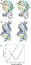
Pseudoknot junctions. (A) A stereo view of the junction between stems S1 (gray) and the triple helix formed by loop L1 (cyan) and stem S2 (orange and yellow). Green lines depict the distance between the O3′ atom at the 3′-end of stem S1 and the O5′ atoms in consecutive residues of the third strand of the triplex. Three green lines correspond to PKs with parameters n1 = 0, 1 and 2. (B) A stereo view of the junction between loop L2 participating in the triplex and the distal end of stem S2. The part of stem S2 not participating in the triplex is shown in blue: the adenine strand in dark blue and the uridine strand in light blue. Green lines show the distance between the O3′ atom at the 3′-end of the third strand of the triplex and the O5′ atoms in consecutive residues of the uridine strand of stem S2. Three green lines correspond to PKs with parameters n2 = 3, 4 and 5. (C) Dependence of the O3′-O5′ distances defined in (A) and (B) on parameters n1 (dashed line) and n2 (solid line). For definition of parameters n1 and n2, see Figure 2A.
Figure 4.
Figure 4.
Ribbon representations of models of telomerase RNA pseudoknots, stereo views. Stem S1 is shown in gray. Residues of stem S2 not participating in base triples are shown in blue. Residues of stem S2 that are part of the triplex are shown in orange (purines) and yellow (pyrimidines). Residues of loop L1, which are part of the triplex, are shown in cyan; the rest of loop L1 is shown in green. Loop L2, if present, is shown in red, and loop L3, in magenta. (A) T. thermophila, (B) T. paravorax, (C) G. chattoni, (D) P. tetraurelia, (E) E. aediculatus and (F) C. colpoda.
Figure 5.
Figure 5.
Structural details of the PK models. The coloring scheme is consistent with that of Figure 4. Filled circles show nitrogen atoms. (A) A stereo view of the interactions of loop L1 residues in the P. tetraurelia PK model, including the uracil participating in the AUU triple and the adenine from junction 1. (B) Unpaired residues of loop L1 (junction 1) of the E. aediculatus form a CpA dinucleotide platform motif. (C) An R-triple GCG from the C. colpoda PK model. (D) A stereo view of an AUA R-triple from the C. colpoda PK model. Note that the loop L1 adenine (label is encircled) forms hydrogen bonds with bases of two neighboring base pairs of stem S2. Ribose and phosphate atoms and the adenine from the upper AU base pair are omitted for clarity. Glycosidic bonds are denoted by ‘Rib’. (E) A stereo view of the network of hydrogen bonds in the C. colpoda PK. Glycosidic bonds are shown in thicker lines. Labels for the loop L1 residues are circled. In the Watson–Crick pairs, only the central N1–N3 hydrogen bonds are shown. The orientation is ∼180° relative to the view in (D).

Similar articles

Cited by

References

    1. Bertuch AA, Lundblad V. The maintenance and masking of chromosome termini. Curr. Opin. Cell Biol. 2006;18:247–253. - PubMed
    1. Autexier C, Lue NF. The structure and function of telomerase reverse transcriptase. Annu. Rev. Biochem. 2006;75:493–517. - PubMed
    1. Romero DP, Blackburn EH. A conserved secondary structure for telomerase RNA. Cell. 1991;67:343–353. - PubMed
    1. Lingner J, Hendrick LL, Cech TR. Telomerase RNAs of different ciliates have a common secondary structure and a permuted template. Genes Dev. 1994;8:1984–1998. - PubMed
    1. Chen JL, Blasco MA, Greider CW. Secondary structure of vertebrate telomerase RNA. Cell. 2000;100:503–514. - PubMed

Publication types