. 2007;35(18):6137-49.
doi: 10.1093/nar/gkm656. Epub 2007 Sep 7.

Tip60 functions as a potential corepressor of KLF4 in regulation of HDC promoter activity

Affiliations

Tip60 functions as a potential corepressor of KLF4 in regulation of HDC promoter activity

Walden Ai et al. Nucleic Acids Res. 2007.

Abstract

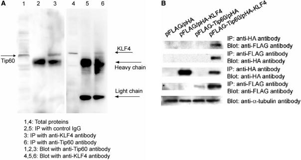
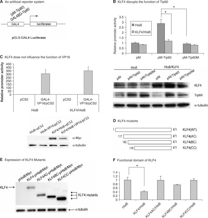
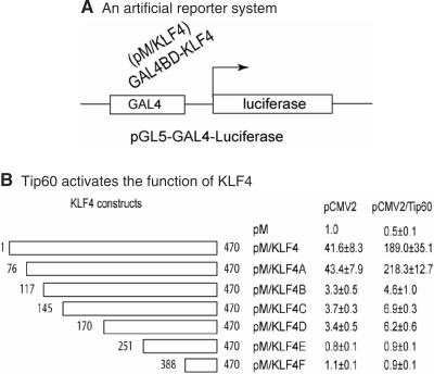
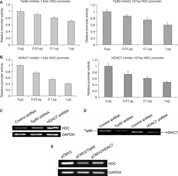
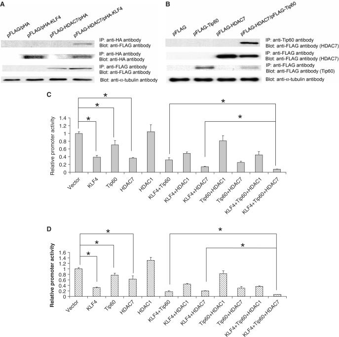
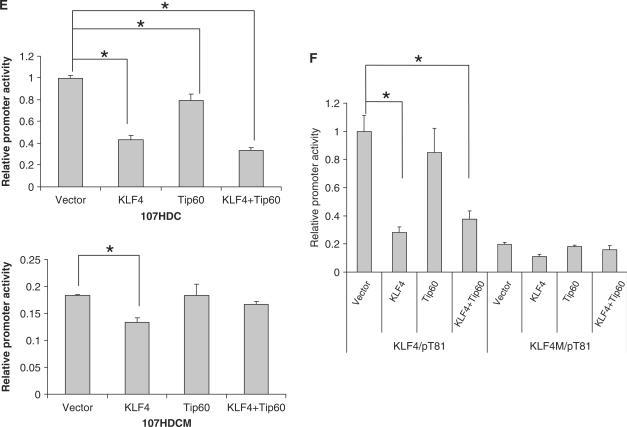
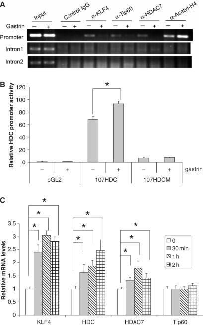
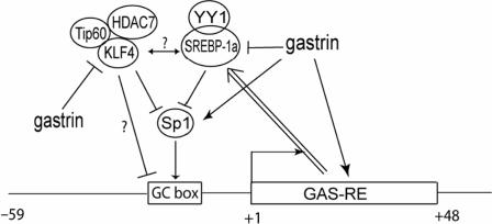
KLF4 is a transcription factor that is highly expressed in the gastrointestinal tract. Previously we have demonstrated that KLF4 represses HDC promoter activity in a gastric cell line through both an upstream Sp1 binding GC box and downstream gastrin responsive elements. However, the mechanism by which KLF4 inhibits HDC promoter is not well defined. In the current study, by using yeast two-hybrid screening, Tip60 was identified as a KLF4 interacting protein. Further coimmunoprecipitation and functional reporter assays support the interaction between these two proteins. In addition, Tip60 and HDAC7, previously shown to interact with each other and repress transcription, inhibited HDC promoter activity in a dose-dependent fashion. Consistently, knock down of Tip60 or HDAC7 gene expression by specific shRNA increased endogenous HDC mRNA level. Co-immunoprecipitation assays showed that HDAC7 was pulled down by KLF4 and Tip60, suggesting that these three proteins form a repressive complex. Further chromatin immuno-precipitation indicated that all three proteins associated with HDC promoter. Two-hour gastrin treatment, known to activate HDC gene expression, significantly decreased the association of KLF4, Tip60 and HDAC7 with HDC promoter, suggesting that gastrin activates HDC gene expression at least partly by decreasing the formation of KLF4/Tip60/HDAC7 repressive complexes at the HDC promoter.


Figures

Figure 1.
Figure 1.
KLF4 interacted with Tip60 by coimmunoprecipitation assay. (A) Endogenous KLF4 interacted with endogenous Tip60. Total proteins were immunoprecipitated with control IgG and anti-KLF4 and anti-Tip60 antibodies. The immunoprecipitates were then probed with anti-Tip60 and anti-KLF4 antibodies. Total proteins were loaded as a control (B). KLF4 interacted with Tip60 in vitro. N-terminally HA-tagged KLF4 expression plasmid (pHA-KLF4) and C-terminally FLAG-tagged Tip60 plasmid (pFLAG-Tip60) with the respective vectors were cotransfected in combination into AGS cells. Total protein extracts were prepared 48 h after transfection. Anti-HA antibody or Anti-FLAG antibody conjugated agarose beads were added to protein extracts. After washing, immunoprecipitated proteins were separated on SDS-PAGE gel and then probed with different antibodies to detect interaction between KLF4 and Tip60.
Figure 2.
Figure 2.
KLF4 disrupts Tip60-mediated transcriptional activation. (A) A reporter system (pGL5-GAL4-Luciferase) was shown where Tip60 was recruited to the proximal promoter region by its Gal4 fusion domain to modulate transcription. (B) KLF4 disrupted Tip60-mediated transcriptional activation. Plasmids expressing KLF4 (KLF4/HisB) and GAL4-fused wild-type and mutant Tip60 (pM-Tip60 and pM-Tip60M) with the respective vectors were cotransfected in combination into AGS cells. Cells were lysed 48 h after transfection followed by luciferase assay as described in Materials and Methods section. Means ± SD for three independent experiments were shown. Statistical difference (P < 0.05) of the relative promoter activities of the transfection experiments was indicated by a star (*). The bottom panel showed the protein levels by western blotting analysis in the same transfection setting using anti-KLF4, anti-Tip60 and anti-α-tubulin antibodies. (C) KLF4 did not influence the function of VP16. As described in panel B, similar transfection experiments were performed using KLF4 and VP16 expressing constructs to detect the effect of KLF4 on VP16-mediated transcriptional activation. The bottom panel showed the protein levels of KLF4 (Myc epitope-tagged) and α-tubulin. (D) Different KLF4 truncation mutants were shown with the starting position of amino acids marked with numbers. (E) Expression of different KLF4 truncation mutants was shown. Total proteins were extracted 48 h after transfection followed by western blotting analysis using anti-Myc antibody. (F) Identification of a functional domain in KLF4. A pM-Tip60 construct was cotransfected with different KLF4 mutant constructs as shown in panel D and the reporter into AGS cells. The relative reporter activities with different KLF4 constructs were measured as described above.
Figure 3.
Figure 3.
Tip60 activates KLF4-mediated transcriptional activation. (A) As described in Figure 2A, a similar artificial promoter system was used where KLF4 was recruited to the proximal promoter region. (B) Tip60 and vector were cotransfected with different pM/KLF4 constructs to test the function of Tip60 in KLF4-mediated transcriptional regulation. The relative promoter activities were normalized with the control KLF4 plasmid (pM) and with control Tip60 plasmid (pCMV2).
Figure 4.
Figure 4.
Tip60 and HDAC7 inhibited HDC promoter activity. (A) Tip60 dose-dependently inhibited both the full length (1.8 kb, left panel) and minimal (107 bp, right panel) HDC promoter. HDC promoter reporter constructs were cotransfected with increasing amount of Tip60 expressing construct into AGS cells. Forty-eight hours after transfection, cells were lysed, luciferase activities were measured and relative promoter activities were calculated as described in Materials and Methods section. (B) HDAC7 dose-dependently inhibited HDC promoter activity. The experiments were performed similarly as described in panel A. In both A and B, the total amount of plasmids in each transfection was 1 μg. The space besides Tip60 or HDAC7 construct was filled with the empty vector. For all these experiments, means ± SD for three independent experiments were shown. (C) Endogenous HDC mRNA levels were elevated by knocking down Tip60 and HDAC7 gene expression. Total RNA was extracted from a control cell line stably expressing a control shRNA, Tip60 shRNA and HDAC7 shRNA stably expressing cell lines. Reverse transcription was performed to generate cDNA. RT-PCR was then followed to amplify a fragment of HDC cDNA and a fragment of GAPDH cDNA. PCR products were separated on agarose gel and visualized under UV light. (D) Expression of Tip60 and HDAC7 in respective cell lines was shown that stably expressed Tip60 or HDAC7 shRNA. RT-PCR was performed as described in panel C to amplify a fragment of Tip60 and HDAC7 cDNA. (E) Endogenous HDC mRNA levels were downregulated by overexpression of exogenous Tip60 and HDAC7. Total RNA was extracted from AGS cells that transfected with vector (pCMV2), Tip60 (pCMV2/Tip60) and HDAC7 (pCMV2/HDAC7). RT-PCR was performed as described above. One representative of each shRNA cell line (control shRNA, Tip60 shRNA and HDAC7 shRNA), and one representative of Tip60 and HDAC7 overexpression experiments was shown from three independent experiments.
Figure 5.
Figure 5.
KLF4, Tip60 and HDAC7 formed a complex and cooperatively inhibited HDC promoter activity. (A) HDAC7 was pulled down by KLF4. Coimmunoprecipitation was performed using anti HA-tag antibody after transient transfection of pHA-KLF4 and pFLAG-HDAC7 constructs into AGS cells as described in Methods and Materials section. Anti Flag-tag antibody was used to detect HDAC7 expression in the following western blotting analysis. (B) HDAC7 was pulled down by Tip60. Similar to A, except anti-Tip60 antibody was used for immunoprecipitation. (C) KLF4, Tip60 and HDAC7 cooperatively repressed 1.8 kb HDC promoter activity. KLF4, Tip60, HDAC7 expressing plasmids with their respective vectors were cotransfected into AGS cells in combination of three constructs with the full length 1.8 kb HDC promoter reporter. Forty-eight hours after transfection, cells were lysed, luciferase activities were measured and relative promoter activities were calculated as described in Materials and Methods section. HDAC1 construct was also used as a control for HDAC7. (D) KLF4, Tip60 and HDAC7 cooperatively repressed 107 bp HDC promoter activity. Similar to panel C except the minimal 107 bp human HDC promoter reporter was used instead of the full length 1.8 kb HDC reporter. (E) Tip60 lost its inhibitory effect when a major KLF4 responsive element (Sp1 binding GC box) was mutated in the HDC promoter. Cotransfection experiments were formed similarly as described above with wild-type 107 bp human HDC promoter reporter (top) and mutant reporter (bottom) with mutations in the upstream GC box. (F) Tip60 did not have any effect on a KLF4 responsive artificial promoter. Cotransfection experiments were formed similarly with an artificial reporter KLF4/pT81 with a KLF4 binding site and various combinations of KLF4 and Tip60 overexpression constructs. A mutant reporter (KLF4M/pT81) with mutations in the KLF4 binding site was also used in the experiment. Means ± SD for three independent experiments were shown for reporter assays (panels C–F), and major statistical difference (P < 0.05) of the relative promoter activities of the transfection experiments was indicated by a star (*).
Figure 5.
Figure 5.
KLF4, Tip60 and HDAC7 formed a complex and cooperatively inhibited HDC promoter activity. (A) HDAC7 was pulled down by KLF4. Coimmunoprecipitation was performed using anti HA-tag antibody after transient transfection of pHA-KLF4 and pFLAG-HDAC7 constructs into AGS cells as described in Methods and Materials section. Anti Flag-tag antibody was used to detect HDAC7 expression in the following western blotting analysis. (B) HDAC7 was pulled down by Tip60. Similar to A, except anti-Tip60 antibody was used for immunoprecipitation. (C) KLF4, Tip60 and HDAC7 cooperatively repressed 1.8 kb HDC promoter activity. KLF4, Tip60, HDAC7 expressing plasmids with their respective vectors were cotransfected into AGS cells in combination of three constructs with the full length 1.8 kb HDC promoter reporter. Forty-eight hours after transfection, cells were lysed, luciferase activities were measured and relative promoter activities were calculated as described in Materials and Methods section. HDAC1 construct was also used as a control for HDAC7. (D) KLF4, Tip60 and HDAC7 cooperatively repressed 107 bp HDC promoter activity. Similar to panel C except the minimal 107 bp human HDC promoter reporter was used instead of the full length 1.8 kb HDC reporter. (E) Tip60 lost its inhibitory effect when a major KLF4 responsive element (Sp1 binding GC box) was mutated in the HDC promoter. Cotransfection experiments were formed similarly as described above with wild-type 107 bp human HDC promoter reporter (top) and mutant reporter (bottom) with mutations in the upstream GC box. (F) Tip60 did not have any effect on a KLF4 responsive artificial promoter. Cotransfection experiments were formed similarly with an artificial reporter KLF4/pT81 with a KLF4 binding site and various combinations of KLF4 and Tip60 overexpression constructs. A mutant reporter (KLF4M/pT81) with mutations in the KLF4 binding site was also used in the experiment. Means ± SD for three independent experiments were shown for reporter assays (panels C–F), and major statistical difference (P < 0.05) of the relative promoter activities of the transfection experiments was indicated by a star (*).
Figure 6.
Figure 6.
KLF4, Tip60 and HDAC7 bind to HDC promoter by chromatin immunoprecipitation assay. (A) Overnight starved AGSE cells were treated with 10−8 M gastrin for 2 h. Cells were then fixed with formaldehyde followed by protein extraction as described in Materials and Methods section. Antibodies against KLF4, Tip60, HDAC7, acetyl-H4 plus control IgG were used to precipitate DNA–protein complexes. Purified DNA samples from precipitated DNA–protein complexes were used as template for PCR to amplify a fragment of HDC promoter, intron 1 and intron 2. (B) Gastrin treatment increased 107HDC promoter activity. Vector (pGL2), 107HDC reporter and 107HDCM reporter were transfected into AGSE cells. Before harvesting, cells were treated with 10−8 M gastrin from 2 h. Then, luciferase assays were performed as described. (C) mRNA levels of KLF4, HDC, Tip60 and HDAC7 after 10−8 M gastrin treatment were shown. Total RNA was extracted after gastrin treatment at different time points. Reverse transcription and follow-up quantitative RT-PCR was performed. Relative mRNA level was calculated as described in Materials and Methods section. Means ± SD for three independent experiments were shown, and statistical difference (P < 0.05) of the relative mRNA levels was indicated by a star (*).
Figure 7.
Figure 7.
A proposed model of transcriptional regulation of HDC is shown by different nuclear factors at the upstream GC box in the promoter. Sp1 has been shown to activate the promoter through this element. Gastrin activates HDC gene expression through both downstream gastrin responsive elements and the upstream GC box. Our published data also suggest that YY1 and SREBP-1a form a complex to compete Sp1 in binding the GC box, resulting in the transcriptional inhibition. The double line with arrow indicates that the downstream GAS-RE is required for this inhibition. Published data and the data presented here suggest that gastrin activates Sp1. Competition between Sp1 and KLF4 on the upstream GC box of the HDC promoter then decreases association of KLF4-Tip60-HDAC7 repressive complexes with the promoter, resulting in the upregulation of HDC gene expression. The relationship between YY1/SREBP-1a complex and KLF4/Tip60/HDAC7 complex remains to be determined.

References

    1. Zhang W, Geiman DE, Shields JM, Dang DT, Mahatan CS, Kaestner KH, Biggs JR, Kraft AS, Yang VW. The gut-enriched Kruppel-like factor (Kruppel-like factor 4) mediates the transactivating effect of p53 on the p21WAF1/Cip1 promoter. J. Biol. Chem. 2000;275:18391–18398. - PMC - PubMed
    1. Garrett-Sinha LA, Eberspaecher H, Seldin MF, de Crombrugghe B. A gene for a novel zinc-finger protein expressed in differentiated epithelial cells and transiently in certain mesenchymal cells. J. Biol. Chem. 1996;271:31384–31390. - PubMed
    1. Yet SF, McA'Nulty MM, Folta SC, Yen HW, Yoshizumi M, Hsieh CM, Layne MD, Chin MT, Wang H, et al. Human EZF, a Kruppel-like zinc finger protein, is expressed in vascular endothelial cells and contains transcriptional activation and repression domains. J. Biol. Chem. 1998;273:1026–1031. - PubMed
    1. Black AR, Black JD, Azizkhan-Clifford J. Sp1 and kruppel-like factor family of transcription factors in cell growth regulation and cancer. J. Cell Physiol. 2001;188:143–160. - PubMed
    1. Chen ZY, Shie JL, Tseng CC. Gut-enriched Kruppel-like factor represses ornithine decarboxylase gene expression and functions as checkpoint regulator in colonic cancer cells. J. Biol. Chem. 2002;277:46831–46839. - PubMed

Publication types

MeSH terms