. 2007;35(17):e116.
doi: 10.1093/nar/gkm466. Epub 2007 Sep 7.

Vitamin H-regulated transgene expression in mammalian cells

Affiliations

Vitamin H-regulated transgene expression in mammalian cells

Wilfried Weber et al. Nucleic Acids Res. 2007.

Abstract

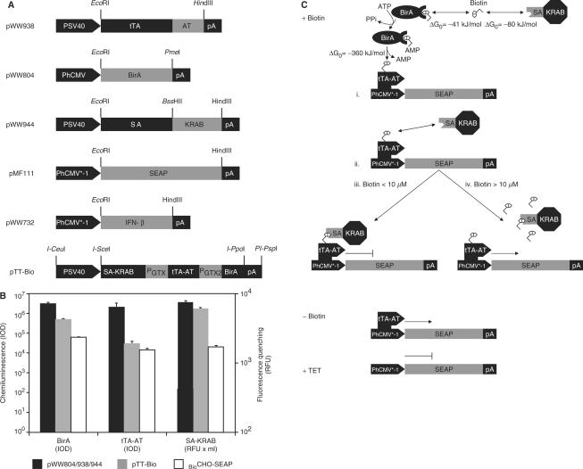
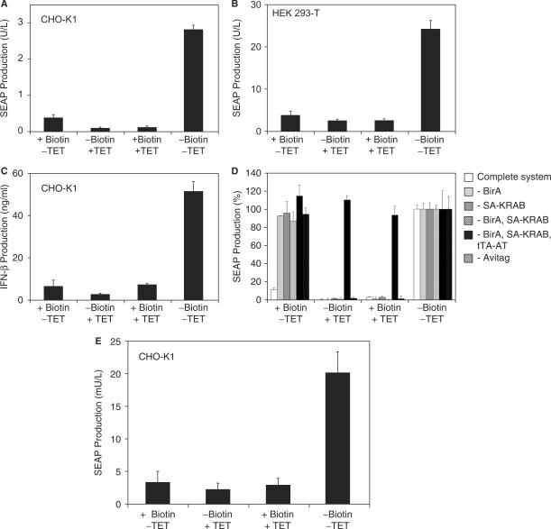
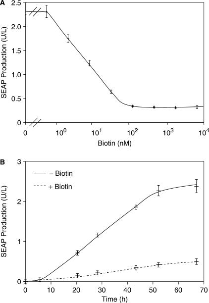
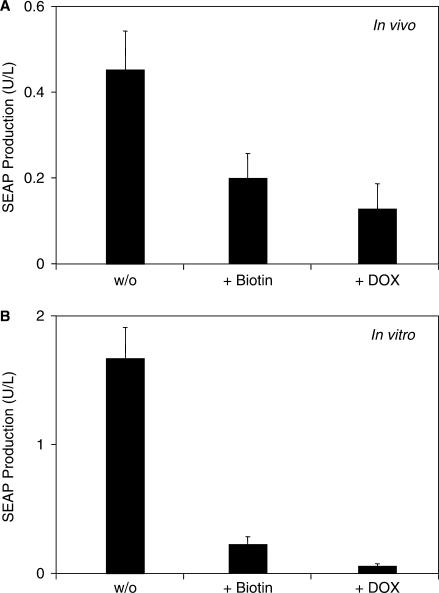
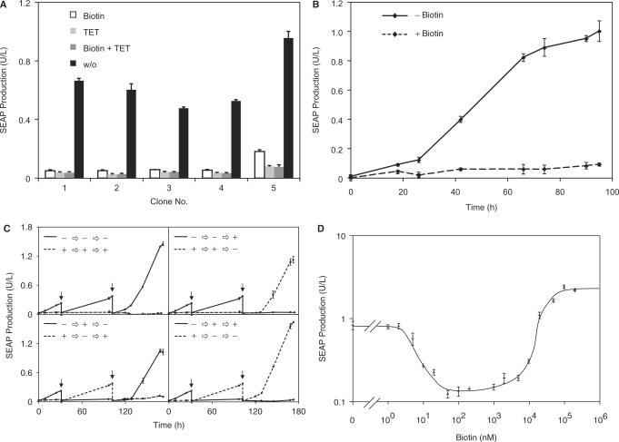
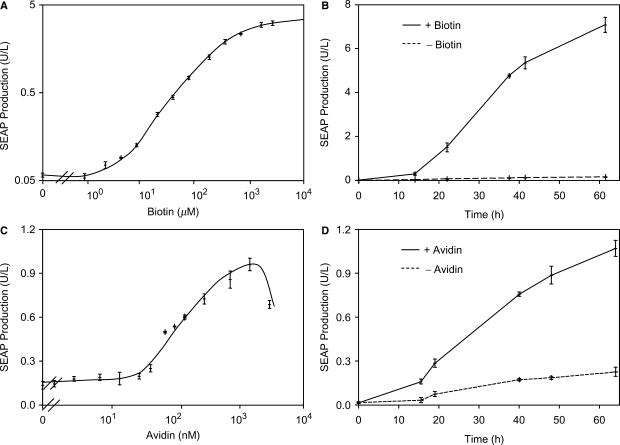
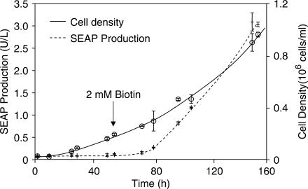
Although adjustable transgene expression systems are considered essential for future therapeutic and biopharmaceutical manufacturing applications, the currently available transcription control modalities all require side-effect-prone inducers such as immunosupressants, hormones and antibiotics for fine-tuning. We have designed a novel mammalian transcription-control system, which is reversibly fine-tuned by non-toxic vitamin H (also referred to as biotin). Ligation of vitamin H, by engineered Escherichia coli biotin ligase (BirA), to a synthetic biotinylation signal fused to the tetracycline-dependent transactivator (tTA), enables heterodimerization of tTA to a streptavidin-linked transrepressor domain (KRAB), thereby abolishing tTA-mediated transactivation of specific target promoters. As heterodimerization of tTA to KRAB is ultimately conditional upon the presence of vitamin H, the system is vitamin H responsive. Transgenic Chinese hamster ovary cells, engineered for vitamin H-responsive gene expression, showed high-level, adjustable and reversible production of a human model glycoprotein in bench-scale culture systems, bioreactor-based biopharmaceutical manufacturing scenarios, and after implantation into mice. The vitamin H-responsive expression systems showed unique band pass filter-like regulation features characterized by high-level expression at low (0-2 nM biotin), maximum repression at intermediate (100-1000 nM biotin), and high-level expression at increased (>100 000 nM biotin) biotin concentrations. Sequential ON-to-OFF-to-ON, ON-to-OFF and OFF-to-ON expression profiles with graded expression transitions can all be achieved by simply increasing the level of a single inducer molecule without exchanging the culture medium. These novel expression characteristics mediated by an FDA-licensed inducer may foster advances in therapeutic cell engineering and manufacturing of difficult-to-produce protein therapeutics.


Figures

Figure 1.
Figure 1.
Vitamin H-responsive gene expression in mammalian cells. (A) Expression vectors for vitamin H-responsive gene expression. AT, synthetic BirA-specific biotinylation signal (Avitag, AT); BirA, Escherichia coli biotin ligase; IFN-β, human beta interferon; PGTX, synthetic promoter; KRAB, krueppel-associated box protein of the human kox-1 gene; pA, simian virus 40-derived polyadenylation signal; PhCMV*−1, tTA-specific tetracycline-responsive promoter; PhCMV, human cytomegalovirus immediate early promoter; PSV40, simian virus 40 promoter; SA, streptavidin; SEAP, human placental secreted alkaline phosphatase; tTA, tetracycline-dependent transactivator. (B) Quantification of BirA, tTA-AT and SA-KRAB expression levels. Cells were transfected with either all three regulatory vectors (pWW804, pWW938 and pWW944) or pTT-Bio alone followed by quantification of the expression levels by chemiluminiscence-based western blot analysis (BirA, tTA-AT) or by incubating SA-KRAB-containing cell lysates with FITC-biotin, the fluorescence of which is quenched upon binding to streptavidin. The expression levels of the three proteins were also quantified in the stable cell line BioCHO-SEAP. IOD, integrated optical density of chemiluminescence signal; RFU, relative fluorescence units. (C) Mode of function. In the presence of biotin (+Biotin), the avitagged tTA (tTA-AT) is biotinylated by BirA (i) thereby triggering binding of streptavidin-KRAB [SA-KRAB, (ii)] and silencing of tTA-AT-mediated PhCMV*−1 activation (iii). At high biotin concentrations all tTA-AT and SA-KRAB-binding sites are saturated thereby preventing heterodimerization and de-repression of the target gene (iv). In the absence of biotin (−Biotin) heterodimerization of tTA-AT and SA-KRAB is prevented and tTA-AT binding to PhCMV*−1 induces SEAP expression. In the presence of tetracycline (+TET), tTA-AT-binding to PhCMV*−1 is prevented regardless of whether or not it has heterodimerized with SA-KRAB, and SEAP expression remains silent. Free standard binding enthalpies (ΔG0) for the covalent biotin-avitag bond as well as for the non-covalent biotin-streptavidin interaction are indicated.
Figure 2.
Figure 2.
Functional validation of vitamin H-responsive expression technology. (A) 40 000 CHO-K1 cells were co-transfected with plasmids pWW944, pWW938, pMF111 and pWW804 (see Figure 1A) and cultivated in biotin-free medium supplemented with (+) or without (−) biotin (100 nM) or tetracycline (TET; 2 µg/ml) for 48 h prior to quantification of SEAP production. (B) 30 000 HEK293-T cells were co-transfected with plasmids pWW944, pWW938, pMF111 and pWW804 (see Figure 1A) and cultivated in biotin-free medium in the presence (+) or absence (−) of exogenous biotin (100 nM) or TET (2 µg/ml) for 48 h prior to quantification of SEAP production. (C) 30 000 CHO-K1 cells were co-transfected with plasmids pWW944, pWW938, pWW732 and pWW804 (see Figure 1A) and cultivated in biotin-free medium in the presence (+) or absence (−) of exogenous biotin (100 nM) or TET (2 µg/ml) for 48 h prior to quantification of beta interferon (IFN-β) production. (D) Specificity of biotin-regulated gene expression. All components of the biotin-responsive expression system [tTA-AT (pWW938), SA-KRAB (pWW944), BirA (pWW804)] were co-transfected with the reporter plasmid pMF111 into 30 000 CHO-K1 which were cultivated for 48 h in the presence (+) or absence (−) of biotin (100 nM) or tetracycline (2 µg/ml) before SEAP production was quantified. In parallel control experiments single components/plasmids were omitted. In order to better visualize the effect of each component, SEAP production was normalized to the inducer-free condition (−Biotin. −TET). (E) Compact genetic design for biotin-regulated gene expression. A total of 30 000 CHO-K1 cells were transfected with plasmids pTT-Bio and pMF111 (see Figure 1A for genotype) and cultivated in the presence (+) or absence (−) of biotin (100 nM) or tetracycline (2 µg/ml) for 48 h prior to quantification of SEAP production.
Figure 3.
Figure 3.
Analysis of vitamin H-responsive transgene expression. (A) Dose-response characteristics. A total of 30 000 CHO-K1 cells were co-transfected with plasmids pWW944, pWW938, pMF111 and pWW804 and cultivated for 48 h in biotin-free medium supplemented with increasing biotin concentrations prior to scoring SEAP production. (B) Expression kinetics. A total of 30 000 CHO-K1 cells were co-transfected with plasmids pWW944, pWW938, pMF111 and pWW804 and SEAP production was profiled for 68 h in the presence (+) or absence (−) of biotin (100 nM).
Figure 4.
Figure 4.
In vivo validation of vitamin H-responsive gene expression. (A) CHO-K1 cells were co-transfected with plasmids pWW944, pWW938, pMF111 and pWW804, encapsulated in coherent alginate-poly-l-lysine-alginate capsules and intraperitoneally injected into biotin-deficient mice (2 × 10⁶ cells/mouse). Mice further received biotin (+Biotin, 100 µg/kg) or doxycycline (+DOX, 100 mg/kg) injections and were subsequently kept for 48 h prior to quantification of SEAP production in the serum. (B) Parallel in vitro validation. Encapsulated CHO-K1 cells co-transfected with plasmids pWW944, pWW938, pMF111 and pWW804 were cultivated in vitro (2 × 10⁶ cells/20 ml biotin-free medium) in the presence of exogenous biotin (+Biotin, 100 nM) or doxycycline (+DOX, 2 µg/ml) for 48 h prior to quantification of SEAP production.
Figure 5.
Figure 5.
Characterization of the transgenic cell line BioCHO-SEAP engineered for vitamin H-responsive SEAP expression. (A) CHO-K1 cells were stably transfected with plasmids pWW944, pWW938, pMF111 and pWW804 (BioCHO-SEAP) and subjected to single-cell cloning. Five representative clones were cultivated (60 000 cells/ml) in biotin-free medium in the presence or absence of biotin (100 nM) or tetracycline (2 µg/ml) for 48 h prior to quantification of SEAP expression. (B) Expression kinetics of BioCHO-SEAP1. SEAP production from BioCHO-SEAP1 cells was monitored when grown in biotin-free medium for 96 h in the presence (+, 100 nM) or absence (−) of biotin. At t = 0 the cells were seeded in the respective media. (C) Reversibility of SEAP expression in BioCHO-SEAP1 cells. SEAP expression from BioCHO-SEAP1 cells was followed during cultivation in biotin-free medium for 170 h in the presence (+) or absence (−) of 100 nM biotin. At the indicated times (arrows) each culture was split into two and cultivated in fresh medium with the same or an alternating −/+ biotin status. (D) Dose-response characteristics of BioCHO-SEAP1 cells. BioCHO-SEAP1 was cultivated (60 000 cells/ml) in biotin-free medium supplemented with increasing biotin concentrations for 48 h prior to quantification of SEAP production.
Figure 6.
Figure 6.
Vitamin H-responsive transgene expression in standard culture environments containing natural biotin levels. (A) Dose-response characteristics of BioCHO-SEAP1 cultivated in standard serum (biotin)-containing medium. BioCHO-SEAP1 (30 000 cells/ml) was cultivated in standard serum (biotin)-containing medium in the presence of increasing biotin concentrations for 48 h prior to scoring SEAP production. (B) Expression kinetics. SEAP production from BioCHO-SEAP1 (60 000 cells/ml) was monitored for 65 h in the presence (+, 2 mM) or absence (−) of biotin supplementation. (C) Regulated SEAP expression by avidin-mediated biotin sequestration. BioCHO-SEAP1 (60 000 cells/ml) was cultivated in the presence of increasing avidin concentrations (expressed as biotin-binding capacity) for 48 h prior to assessing SEAP production. (D) Avidin-controlled induction kinetics of vitamin H-regulated SEAP expression. SEAP production from BioCHO-SEAP1 cells (60 000 cells/ml) was monitored during cultivation for 65 h in the presence (+, 1.5 µM) or absence (−) of avidin.
Figure 7.
Figure 7.
Performance of vitamin H-responsive gene expression during cultivation in bioreactors. BioCHO-SEAP1 was cultivated in a BioWave® bioreactor in 1 l culture volume using serum- (biotin) containing standard medium. SEAP expression and cell density were monitored over time. After 53 h (arrow) SEAP expression was induced by adding 2 mM biotin.

Similar articles

Cited by

References

    1. Kramer BP, Fischer C, Fussenegger M. BioLogic gates enable logical transcription control in mammalian cells. Biotechnol. Bioeng. 2004;87:478–484. - PubMed
    1. Kramer BP, Fussenegger M. Hysteresis in a synthetic mammalian gene network. Proc. Natl Acad. Sci. USA. 2005;102:9517–9522. - PMC - PubMed
    1. Kramer BP, Viretta AU, Daoud-El-Baba M, Aubel D, Weber W, Fussenegger M. An engineered epigenetic transgene switch in mammalian cells. Nat. Biotechnol. 2004;22:867–870. - PubMed
    1. Kramer BP, Weber W, Fussenegger M. Artificial regulatory networks and cascades for discrete multilevel transgene control in mammalian cells. Biotechnol. Bioeng. 2003;83:810–820. - PubMed
    1. McLoughlin JM, McCarty TM, Cunningham C, Clark V, Senzer N, Nemunaitis J, Kuhn JA. TNFerade, an adenovector carrying the transgene for human tumor necrosis factor alpha, for patients with advanced solid tumors: surgical experience and long-term follow-up. Ann. Surg. Oncol. 2005;12:825–830. - PubMed

Publication types

MeSH terms