. 2007 Dec;56(12):2919-26.
doi: 10.2337/db07-0773. Epub 2007 Sep 7.

Aberrant endometrial features of pregnancy in diabetic NOD mice

Affiliations

Aberrant endometrial features of pregnancy in diabetic NOD mice

Suzanne D Burke et al. Diabetes. 2007 Dec.

Abstract

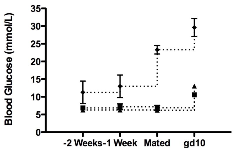
Objective: Pregnant diabetic women are at a 4-12 times higher risk for preeclampsia, an urgent acute-onset complication of mid- to late gestation, than normal pregnant women. Hallmarks of preeclampsia are hypertension, proteinuria, and incomplete modification of endometrial spiral arteries. Transient proangiogenic lymphocytes called uterine natural killer (uNK) cells are implicated in human and rodent spiral artery modification. We studied mid- to late gestations in spontaneously type 1 diabetic NOD mice to investigate whether diabetes alters uNK cell homing and/or function.

Research design and methods: Normoglycemic, pre-diabetic, and diabetic NOD mice and controls were mated. Lymphocytes and endometrial endothelium and decidua were studied histologically and in functional assays.

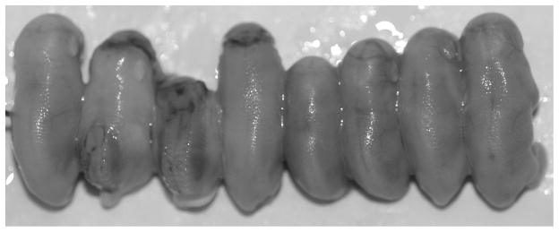
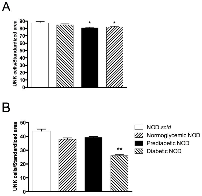
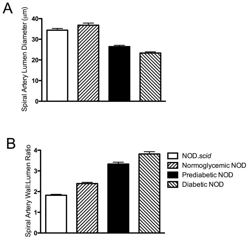
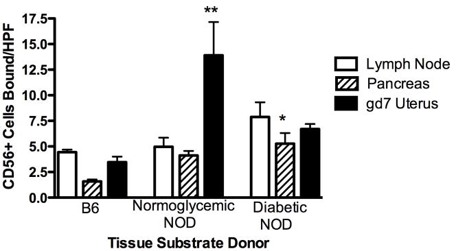
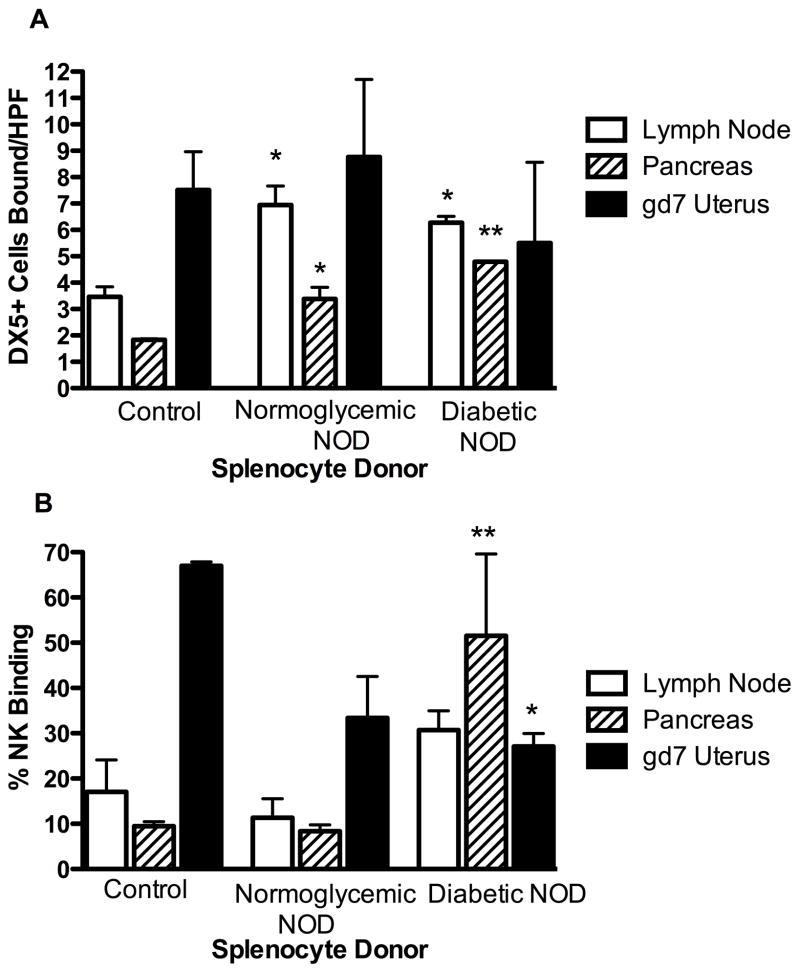
Results: Conception accelerated progression to overt diabetes in NOD females who had limited spiral artery development, heavier placentas, and lighter fetuses displaying numerous birth defects compared with controls. UNK cell numbers were reduced in the decidua basalis of diabetic females, whereas interferon-gamma production was elevated. In diabetic NOD mice, decidual expression of the mucosal vascular addressin cell adhesion molecule (MAdCAM)-1 was aberrant in position, whereas vascular cell adhesion molecule (VCAM)-1 expression was reduced. Assays of lymphocyte adhesion to tissue sections under shear forces indicated that diabetes compromises the potential homing functions of both endometrial endothelium and peripheral NK cells.

Conclusions: In diabetes, gestational endometrium has immune and vascular defects that likely contribute to murine fetal loss and birth defects. Analogous problems and preeclampsia in diabetic women may involve similar mechanisms.


Figures

Figure 1
Figure 1. Blood glucose values of mated NOD mice
Mice were classified as normoglycemic (n=5; squares; <11 mmol/L), prediabetic (n=7; triangles; >11.1 – 14.9 mmol/L), or diabetic (n=7; diamonds; >15 mmol/L) based on values measured at gd10. Pregnancy elevated blood glucose values in all animals with the largest increase in diabetic mice before conception. Diabetic NOD mice showed an increase in blood glucose from three previous measurements (p<0.005).
Figure 2
Figure 2. Litter of gd18 fetuses born to overtly diabetic NOD dam
Three neural tube defects and widely variable fetal size were noted. At necropsy only 50% had the potential to survive; all exhibited congenital defects.
Figure 3
Figure 3. UNK cell counts within the A) MLAp and B) decidua basalis of NOD.scid, normoglycemic, prediabetic and diabetic NOD implantation sites
*p<0.05 compared to NOD.scid implantation sites. **p<0.001 compared to normoglycemic and prediabetic NOD implantation sites. Data are mean ± SEM from n=3 mice per group (3 viable implantation sites per mouse).
Figure 4
Figure 4. A) Spiral artery lumen diameter (μm) in NOD.scid, normoglycemic, prediabetic, and diabetic NOD mice at gd10. B) Wall:lumen ratios of spiral arteries in NOD.scid, normoglycemic, prediabetic, and diabetic implantation sites
All groups are significantly different, p<0.05, except for NOD.scid vs. normoglycemic NOD mice. Data are mean ± SEM from n=3 mice per group (3 viable implantation sites per mouse).
Figure 5
Figure 5. Representative VCAM-1 staining in gd8 mouse tissues
Endothelium in vessels of decidua basalis consistently appeared more reactive in B6 mice (arrow) A), than in diabetic NOD mice (arrow) B). Pancreatic islets of B6 mice were unreactive C), but were strongly reactive in diabetic NOD mice (arrow) D). The latter showed leukocyte infiltration (arrowhead), consistent with insulitis observed in overt diabetes. Tissues illustrated are from paired single donors. Counterstained with Hematoxylin. Bar = 20μm.
Figure 6
Figure 6. Human CD56+ cells bound to peripheral lymph node (LN) from B6, normoglycemic NOD, prediabetic NOD, or diabetic NOD mice per high-powered field (400× magnification)
Data are presented from replicate experiments using the same blood donor. *p<0.05 vs. B6, **p<0.05 vs. B6 and diabetic NOD mice.
Figure 7
Figure 7. A) FITC-DX5+ splenic NK cells or B) DX5+/CMAC+ splenic NK/lymphocytes bound to peripheral lymph node (LN), pancreas, or gd7 uterus from normal B6 mice per high-powered field (400× magnification)
Splenocyte donors are B6, normoglycemic NOD, or diabetic NOD mice. *p<0.05 vs. B6, **p<0.05 vs. B6 and normoglycemic NOD splenocyte binding.

Similar articles

Cited by

References

    1. Leiter EH. The NOD mouse: a model for insulin-dependent diabetes mellitis. Current Protocols in Immunology. 1997;24(suppl):15.9.1–15.9.23. - PubMed
    1. Wicker LS, Clark J, Fraser HI, Garner VE, Gonzalez-Munoz A, Healy B, Howlett S, Hunter K, Rainbow D, Rosa RL, Smink LJ, Todd JA, Peterson LB. Type 1 diabetes genes and pathways shared by humans and NOD mice. J Autoimmun. 2005;25 (Suppl):29–33. - PubMed
    1. Wicker LS, Todd JA, Peterson LB. Genetic control of autoimmune diabetes in the NOD mouse. Annu Rev Immunol. 1995;13:179–200. - PubMed
    1. Hanninen A, Taylor C, Streeter PR, Stark LS, Sarte JM, Shizuru JA, Simell O, Michie SA. Vascular addressins are induced on islet vessels during insulitis in nonobese diabetic mice and are involved in lymphoid cell binding to islet endothelium. J Clin Invest. 1993;92:2509–2515. - PMC - PubMed
    1. Serreze DV, Leiter EH. Defective activation of T suppressor cell function in nonobese diabetic mice. Potential relation to cytokine deficiencies. J Immunol. 1988;140:3801–3807. - PubMed

Publication types

Grants and funding