. 2007 Nov;152(6):880-90.
doi: 10.1038/sj.bjp.0707449. Epub 2007 Sep 10.

Failure of antioxidants to protect against angiotensin II-induced aortic rupture in aged apolipoprotein(E)-deficient mice

Affiliations

Failure of antioxidants to protect against angiotensin II-induced aortic rupture in aged apolipoprotein(E)-deficient mice

F Jiang et al. Br J Pharmacol. 2007 Nov.

Abstract

Background and purpose: Oxidative stress may be involved in the development of abdominal aortic aneurysms (AAAs). Previous studies indicate that antioxidants protect against AAA formation during chronic angiotensin (Ang) II infusion in apolipoprotein E-deficient (ApoE(0)) mice. We here examine if these protective effects also occurred in aged ApoE(0) mice.

Experimental approach: Male ApoE(0) mice (50-60 weeks) were randomly divided into 4 groups: saline, Ang II (1000 ng kg(-1) min(-1) for 4 weeks), Ang II plus antioxidants (0.1% vitamin E in food plus 0.1% vitamin C in drinking water), and Ang II plus losartan (30 mg kg(-1) day(-1)).

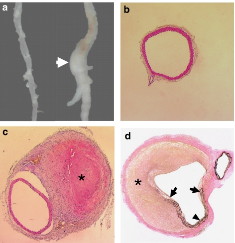
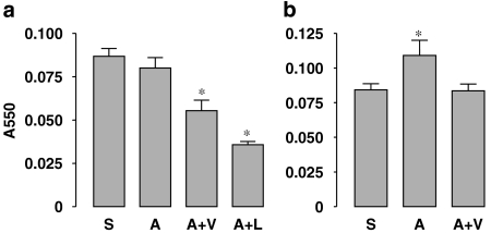
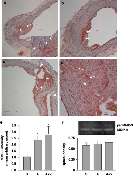
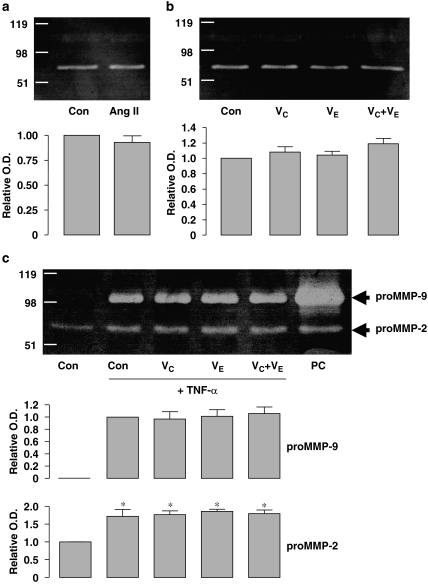
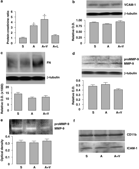
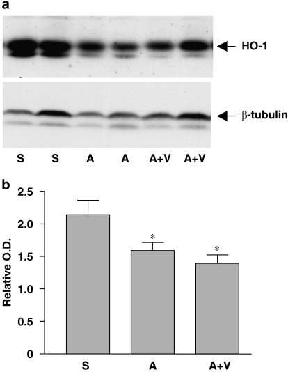
Key results: Exogenous Ang II increased systolic blood pressure by 40 mmHg and resulted in the formation of pseudoaneurysms (rupture and extramural haematoma) in the abdominal aorta in 50% of animals. True aneurysmal dilatation was rarely observed. Antioxidants decreased systemic oxidative stress (plasma malondialdehyde), but had only minor effects on aortic rupture, relative to the complete prevention by losartan. Immunohistochemistry revealed strong matrix metalloproteinase-9 (MMP-9) expression in atherosclerotic plaques and at the sites of rupture. Antioxidants did not affect tumour necrosis factor-alpha-stimulated MMP-9 release from U937 cells. In addition, antioxidants had little effects on Ang II-induced renal dysfunction.

Conclusions and implications: In contrast to previous findings in younger mice, antioxidants had only minor effects on Ang II-induced aortic rupture in aged mice. Our results demonstrate that the pathological features of the aneurysmal remodelling induced by Ang II in old ApoE(0) mice are distinct from those of human AAA.


Figures

Figure 1
Figure 1
Histopathology of Ang II infusion-induced pseudoaneurysm in the abdominal aorta of aged ApoE0 mice. (a) Gross appearance of the pseudoaneurysm (arrow). The aorta from saline treated animal was shown on the left as control. (b) Haematoxylin andeosin (H&E) staining of the transverse section of the suprarenal abdominal aorta from the control group (× 40). (c) Transverse section (× 40) cut from the segment immediately adjacent to the rupture site showing the intact aortic wall. True aneurysmal expansion of the aorta could not be identified. *A thrombus encapsulated in fibrous tissues (H&E staining). (d) Verhoeff's von Giessen staining showing the total rupture of the aortic wall (arrows). A pseudolumen was formed by the thrombus (*) and the residual vessel wall (arrowhead) (× 40). Ang II, Angiotensin II; ApoE0, apolipoprotein E-deficient.
Figure 2
Figure 2
Effects of Ang II and antioxidants on the malondialdehyde (MDA) levels in the plasma (a) and kidney homogenates (b) measured by the thiobarbituric acid-reactive substances (TBARS) assay. S, saline; A, Ang II; V, antioxidant vitamins; L, losartan. Data are mean±s.e.mean. *P<0.05 vs saline, n=5–9. Ang II, Angiotensin II.
Figure 3
Figure 3
Matrix metalloproteinase-9 (MMP-9) production in the aorta examined by immunohistochemistry (ad) and in the plasma examined by gelatin zymography (f). Strong MMP-9 immunoreactivity was identified within atherosclerotic plaques (arrows) in both saline- (a) and Ang II-treated (b) aortas. The inset in panel (a) shows that pre-existing elastic lamina disruptions (arrowheads) can be found underneath the plaque before Ang II treatment. (c) In some specimens from the Ang II group, total medial breakage (arrowheads) in association with localized MMP-9 expression (arrows) was observed before haematoma formation, which may represent a precursor of the pseudoaneurysm. (d) MMP-9 was also highly expressed in the thrombi (*) inside the sac of pseudoaneurysms. Scale bars=50 μm (ac) and 20 μm (d). (e) Quantitation of the intensity of MMP-9 immunoreactivity in the non-ruptured part of the suprarenal abdominal aorta using an arbitrary score (S, saline; A, Ang II; V, antioxidant vitamins; data are mean±s.e.mean. *P<0.05 vs saline, n=6–9). (f) Gelatin zymography and quantitative optical density data of the (pro)MMP-9 activity in the plasma (mean±s.e.mean. n=5–6). Ang II, Angiotensin II.
Figure 4
Figure 4
Effects of Ang II, TNF-α and antioxidant vitamins on matrix metalloproteinase (MMP) release from cultured U937 mononuclear cells. (a and b) Gelatin zymography showing the MMP activity of the conditioned media after a 24-h treatment with vehicle (Con), Ang II (100 nM), vitamin C (VC, 1 mM), vitamin E (VE, 10 μM) or VC+VE. The bands at ∼70 kDa represent the proMMP-2 activity. MMP-9 activity could not be detected under these conditions. Quantitative optical density data are shown below (n=3). (c) Zymography showing that TNF-α (20 ng ml−1 for 24 h) induced proMMP-9 (∼98 kDa) release and enhanced the proMMP-2 activity from U937 cells. Co-treatment with antioxidants had no effect on TNF-α-induced MMP production. *P<0.05 vs control (Con), n=3. Cells treated with phorbol 12-myristate 13-acetate (PMA, 10 ng ml−1) was used as positive control (PC). Ang II, Angiotensin II.
Figure 5
Figure 5
Effects of Ang II and antioxidant co-treatment on renal parameters. (a) Protein to creatinine ratio measured in urine samples from different groups. *P<0.05 vs S, n=5–7. (bd) Western blot analyses for the expression of VCAM-1, fibronectin (FN) and (pro)MMP-9 in the total protein extracts of kidney. The level of β-tubulin was used to normalize the protein load. Quantitative densitometry data (target to β-tubulin ratio) are shown below each blot. (e) Gelatin zymography and quantitative optical density data of the (pro)MMP-9 activity in kidney homogenates. (f) Western blots of CD11b and ICAM-1 expression in the kidney. Example from two separate experiments. S, saline; A, Ang II; V, antioxidant vitamins; L, losartan. Ang II, Angiotensin II; ICAM-1, intercellular adhesion molecule-1; VCAM-1, vascular cell adhesion molecule-1.
Figure 6
Figure 6
Expression of HO-1 in kidneys from saline (S)-, Ang II (A)- and Ang II plus antioxidant vitamins (A+V)-treated mice. (a) Western blot of HO-1. (b) Quantitation of the western blot by densitometry. Data are expressed as HO-1 to β-tubulin ratio. *P<0.05 vs S, n=9–11. Ang II, Angiotensin II; HO-1, haeme oxygenase-1.

References

    1. Agarwal R, Campbell RC, Warnock DG. Oxidative stress in hypertension and chronic kidney disease: role of angiotensin II. Semin Nephrol. 2004;24:101–114. - PubMed
    1. Aizawa T, Ishizaka N, Taguchi J, Nagai R, Mori I, Tang SS, et al. Heme oxygenase-1 is upregulated in the kidney of angiotensin II-induced hypertensive rats: possible role in renoprotection. Hypertension. 2000;35:800–806. - PubMed
    1. Araujo M, Welch WJ. Oxidative stress and nitric oxide in kidney function. Curr Opin Nephrol Hypertens. 2006;15:72–77. - PubMed
    1. Arenas IA, Xu Y, Lopez-Jaramillo P, Davidge ST. Angiotensin II-induced MMP-2 release from endothelial cells is mediated by TNF-alpha. Am J Physiol Cell Physiol. 2004;286:C779–C784. - PubMed
    1. Bellosta S, Via D, Canavesi M, Pfister P, Fumagalli R, Paoletti R, et al. HMG-CoA reductase inhibitors reduce MMP-9 secretion by macrophages. Arterioscler Thromb Vasc Biol. 1998;18:1671–1678. - PubMed

Publication types

MeSH terms