. 2007 Sep 28;13(36):4881-90.
doi: 10.3748/wjg.v13.i36.4881.

Impaired contractility and remodeling of the upper gastrointestinal tract in diabetes mellitus type-1

Affiliations

Impaired contractility and remodeling of the upper gastrointestinal tract in diabetes mellitus type-1

Jens Brondum Frokjaer et al. World J Gastroenterol. .

Abstract

Aim: To investigate that both the neuronal function of the contractile system and structural apparatus of the gastrointestinal tract are affected in patients with longstanding diabetes and auto mic neuropathy.

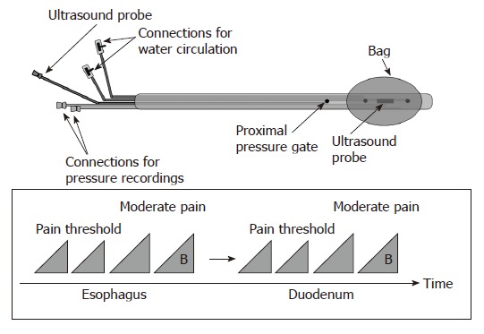
Methods: The evoked esophageal and duodenal contractile activity to standardized bag distension was assessed using a specialized ultrasound-based probe. Twelve type-1 diabetic patients with autonomic neuropathy and severe gastrointestinal symptoms and 12 healthy controls were studied. The geometry and biomechanical parameters (strain, tension/stress, and stiffness) were assessed.

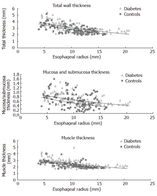
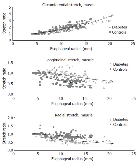
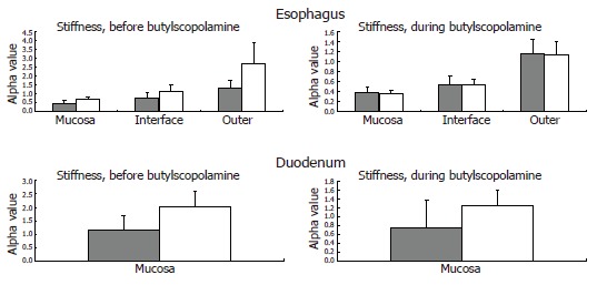
Results: The diabetic patients had increased frequency of distension-induced contractions (6.0 +/- 0.6 vs 3.3 +/- 0.5, P < 0.001). This increased reactivity was correlated with the duration of the disease (P = 0.009). Impaired coordination of the contractile activity in diabetic patients was demonstrated as imbalance between the time required to evoke the first contraction at the distension site and proximal to it (1.5 +/- 0.6 vs 0.5 +/- 0.1, P = 0.03). The esophageal wall and especially the mucosa-submucosa layer had increased thickness in the patients (P < 0.001), and the longitudinal and radial compressive stretch was less in diabetics (P < 0.001). The esophageal and duodenal wall stiffness and circumferential deformation induced by the distensions were not affected in the patients (all P > 0.14).

Conclusion: The impaired contractile activity with an imbalance in the distension-induced contractions likely reflects neuronal abnormalities due to autonomic neuropathy. However, structural changes and remodeling of the gastrointestinal tract are also evident and may add to the neuronal changes. This may contribute to the pathophysiology of diabetic gut dysfunction and impact on future management of diabetic patients with gastrointestinal symptoms.


Figures

Figure 1
Figure 1
The probe design allows bag distension of the esophagus and duodenum together with cross-sectional ultrasound imaging and recording of the bag and proximal pressures (top). The distension protocol is shown at the bottom. The first distensions to the pain threshold were preconditioning stimuli also used for learning. They were followed by a distension (15 mL/min in the esophagus and 25 mL/min in the duodenum) to the perception of moderate pain. Finally, a bolus of 20 mg of butylscopolamine (B) was given intravenously in order to diminish distension-evoked smooth muscle contractions and a final distension to the perception of moderate pain was done.
Figure 2
Figure 2
Schematic illustration of the neural pathways involved in distension-induced contraction. The primary afferent neurons, the excitatory interneurons and efferent neurons are shown. The dotted neurons illustrate extrinsic pathways. The induction of contraction on the bag depends on the local reflex arc (bottom). The measurement 6 cm proximal to the bag depends on a longer reflex arc including extrinsic pathways (top).
Figure 3
Figure 3
The cross-sectional ultrasound image of the distended distal esophagus allows identification of the esophageal layers, i.e. mucosa-submucosa, muscle and adventitia layers. The white round shadow in the centre is caused by the intraluminal ultrasound probe.
Figure 4
Figure 4
The distension-induced change in thicknesses of the total esophageal wall structure, the muscle layer, and mucosa-submucosa layer during smooth muscle relaxation with butylscopolamine are illustrated as function of the esophageal radius. The data points represent the multiple measuring points during each distension of the patients and controls. Exponential trend lines (solid lines) of the patients and controls are shown. The total wall thickness and the mucosa-submucosa layer were increased in the diabetic patients.
Figure 5
Figure 5
The distension-induced change in circumferential, longitudinal and radial stretch ratios are illustrated as function of the esophageal radius. The curves were obtained during smooth muscle relaxation with butylscopolamine. The data points represent the multiple measuring points during each distension of the patients and controls. Exponential trend lines (solid lines) of the patients and controls are shown. The shortening during distension was clearly reduced in the diabetic patients while the radial stretch was decreased.
Figure 6
Figure 6
The computed esophageal stiffness (circumferential [alpha] constant) of the mucosa, submucosa-muscle interface and outer surface are illustrated. The solid grey bars represent the controls, while the grey-white bars represent the diabetic patients. The stiffness tends to be higher in the diabetic patients and is normalized during smooth muscle relaxation with butylscopolamine. This indicates an increased esophageal resting tone in diabetes. The computed duodenal stiffness is also illustrated. Mean and SEM values are shown.
Math 1
Math 1
Math(A1).
Math 2
Math 2
Math(A1).
Math 3
Math 3
Math(A1).
Math 4
Math 4
Math(A1).
Math 5
Math 5
Math(A1).
Math 6
Math 6
Math(A1).

References

    1. Rundles RW. Diabetic Neuropathy. Medicine. 1945;24:111–160.
    1. Ko GT, Chan WB, Chan JC, Tsang LW, Cockram CS. Gastrointestinal symptoms in Chinese patients with Type 2 diabetes mellitus. Diabet Med. 1999;16:670–674. - PubMed
    1. Folwaczny C, Riepl R, Tschöp M, Landgraf R. Gastrointestinal involvement in patients with diabetes mellitus: Part I (first of two parts). Epidemiology, pathophysiology, clinical findings. Z Gastroenterol. 1999;37:803–815. - PubMed
    1. Spångéus A, El-Salhy M, Suhr O, Eriksson J, Lithner F. Prevalence of gastrointestinal symptoms in young and middle-aged diabetic patients. Scand J Gastroenterol. 1999;34:1196–1202. - PubMed
    1. Horowitz M, Samsom M. Gastrointestinal Function in Diabetes Mellius. Chichester: John Wiley & Sons Ltd; 2004. pp. 1–27.

Publication types