. 2007 Oct 9;46(40):11240-52.
doi: 10.1021/bi700580y. Epub 2007 Sep 11.

Sequence-specific triple helix formation with genomic DNA

Affiliations

Sequence-specific triple helix formation with genomic DNA

Zhaoyang Ye et al. Biochemistry. .

Abstract

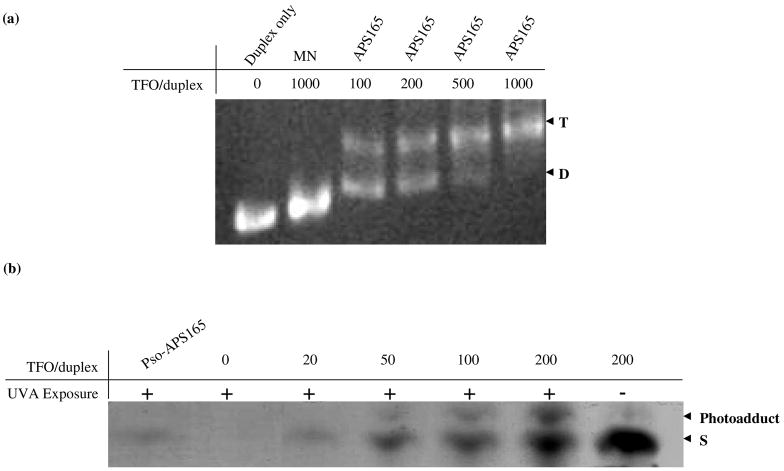
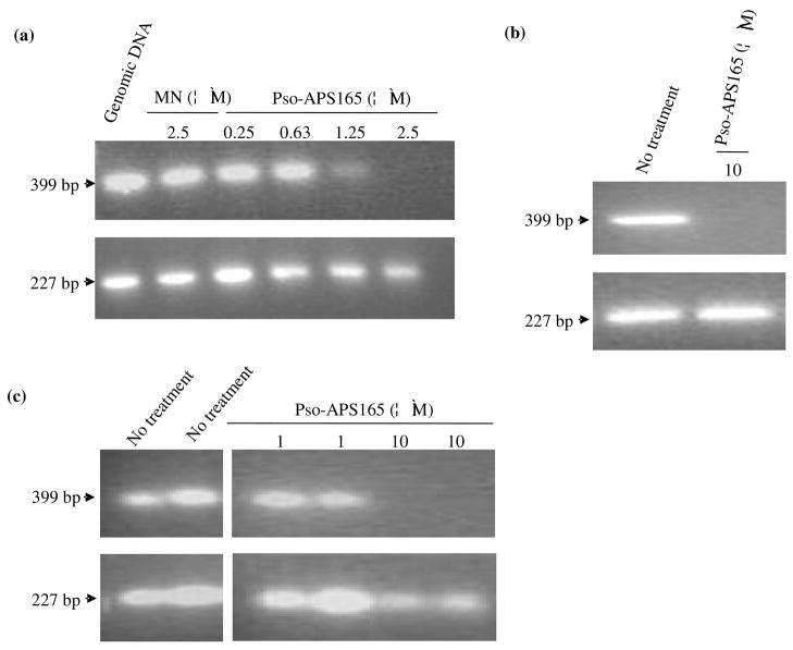
We have previously demonstrated site-specific delivery of antiparallel phosphorothioate triplex forming oligonucleotide (TFO) specific to -165 to -141 promoter region of alpha1(I) collagen (abbreviated as APS165) to hepatic stellate cells (HSCs) of fibrotic rats after conjugation with mannose 6-phosphate-bovine serum albumin. However, we still need to determine whether there is correlation between transcription inhibition and triplex formation with genomic DNA. In this study, APS165 was modified with psoralen and the extent of triplex formation with alpha1(I) collagen DNA was determined in naked genomic DNA, isolated nuclei of HSC-T6 cells and whole cells by using a simple real-time PCR based method. In this method, a purification step was added to remove unbound APS165, which eliminated the possible artifacts during real-time PCR. Psoralen photoadduct formation was shown to be essential to retain triplex structure under denaturing conditions. On naked genomic DNA, 82.2% of DNA formed triplex and 36.7% of genomic DNA in isolated nuclei at 90 min contained triplex structure. As quantified by real-time PCR, 50% of genomic DNA in living cells at 12 h postincubation contained triplex structures. Furthermore, the triplex formation was dose-dependent with 26.5% and 50% of DNA having triplex structure at concentrations of 1 microM and 5 microM, respectively. Moreover, on a plasmid pCol-CAT220 containing rat alpha1(I) gene promoter (-225 to +113), 75.3% of triplex formation was observed, which was correlated with a 73.6% of transcription inhibition. These findings will further strengthen the therapeutic applications of APS165.


Figures

Figure 1
Figure 1
Target sequence and triplex forming oligonucleotides. (a) The promoter region of rat α1(I) collagen gene was shown. The triplex formation site of APS165 in the gene sequence (corresponding to positions between −165 and −141) was indicated. T1 and T2 are 30-nt oligonucleotides overlapping triplex formation site (underlined). Sequences of the triplex forming oligonucleotide APS165 and the control oligonucleotide MN (mismatched sequence, mismatched nucleotides highlighted in italics) are shown. Fluorescein (F) and psoralen (Pso) modifications in the sequences are indicated. APS165 is a 25-nt oligopurine and anti-parallel to the target gene sequence. In addition, APS165 and MN are phosphorothioate modified oligonucleotides. P1–P6, positions of PCR-primers and the lengths of the PCR amplified fragments are indicated for the corresponding set of primers. (b) pCol-CAT220 containing a 338-bp fragment of the rat α1(I) collagen gene (−225 to +113) was shown with BglII, BseRI and XbaI restriction sites. Primers (P2 and P6-P8) were illustrated.
Figure 2
Figure 2
a) Effect of TFO/pDNA ratio on triplex formation. pCol-CAT220 was incubated with Pso-APS165 at 300-fold molar excess. Following UV irradiation, the mixture was purified, and digested with BglII, BseRI and XbaI. The lengths of digested products (64bp, 166 bp and 108 bp) are indicated. Inhibition of cleavage by BseRI resulted in the disappearance of the 108-bp fragment and appearance of a new band of 172bp+25nt of APS165. b) Time course of the reaction of Pso-APS165 with pCol-CAT220. pCol-CAT220 was incubated with Pso-TFO165 at 300-fold molar excess for 90 min at 37 °C, followed by UVA irradiation for 0, 1 and 10 min. The plasmid was then digested with XbaI and BglII to release a 338bp fragment. The samples were finally applied to denaturing gel electrophoresis.
Figure 3
Figure 3
Triplex formation between APS165 with C1 duplex DNA. a) Effect of TFO/duplex DNA ratio on triplex formation. The formation of duplex and triplex was checked by electrophoresis on a native polyacrylamide gel. b) Effect of TFO/duplex ratio and UVA exposure on psoralen-induced photoadduct formation between Pso-APS165 and duplex DNA. Irradiation was carried out for 10 min at 0 °C at 366 nm. Photoadduct was checked by electrophoresis on a 15% denaturing polyacrylamide gel containing 7M urea and 40% formamide.
Figure 4
Figure 4
Triplex formation with genomic DNA, isolated nuclei and living cells. a) Genomic DNA was incubated with Pso-APS165 or control oligonucleotide MN. Samples were then UVA irradiated for 10 min and then subjected to PCR reaction without any manipulation. b) Isolated nuclei were incubated with or without 10 μM of Pso-APS165 for 90 min at 37 °C with gentle rotation, and UVA irradiated at 0 °C. The nuclei were then digested and DNA was isolated. The isolated DNA (2 μg) was subjected to PCR amplification using primers P1 and P2. c) HSC-T6 cells were incubated with or without Pso-APS165 at 37 °C for 12 h, UVA irradiated and the cells were harvested to isolate genomic DNA, which was subjected to PCR reactions. The PCR products were analyzed by 2% agarose gel electrophoresis.
Figure 5
Figure 5
Quantitative measurement of triplex formation in vitro. a) pCol-CAT220 (1μg) was incubated with different amounts of Pso-APS165. Samples were then UVA irradiated for 10 min and then the mixture was subjected to 0.5% agarose gel elution and extraction. Aliquots were subjected to real-time PCR. b) Genomic DNA (4μg) was incubated with different amounts of Pso-APS165. Samples were then UVA irradiated for 10 min and then the mixture was subjected to 0.5% agarose gel elution and extraction. Aliquots were subjected to real-time PCR. The amounts of PCR products of target fragments were calculated based on difference between Ct numbers obtained for the same sample from a control region and target region. Relative PCR product was reported as a function of Pso-APS165 concentration by setting sample without TFO treatment as 100%. Data were expressed as the mean ± standard deviation (SD) (n = 3). *: P < 0.05 was considered statistically significant. Statistics were calculated based on unpaired student’s t-test. (+: with UVA irradiation; −: without UVA irradiation)
Figure 6
Figure 6
Quantitative measurement of triplex formation in nuclei and living cells. a) Isolated nuclei were incubated with Pso-APS165 at concentrations of 0, 5 and 10 μM for 90 min at 37 °C. The nuclei were then UVA irradiated for 10 min and then genomic DNA was isolated and purified. Aliquots of DNA samples were subjected to real-time PCR. b) HSC-T6 cells were incubated with Pso-APS165 or APS165 at concentrations of 0, 1 and 5 μM at 37 °C for 12 h, UVA irradiated and the cells were harvested to isolate genomic DNA. The DNA was purified by gel elution and aliquots were subjected to real-time PCR. The amounts of PCR products of target fragments were calculated based on difference between Ct numbers obtained for the same sample from a control region and target region. Relative PCR product was reported as a function of Pso-APS165 concentration by setting sample without TFO treatment as 100%. Data were expressed as the mean ± standard deviation (SD) (n = 3). *: P < 0.05 was considered statistically significant. Statistics were calculated based on unpaired student’s t-test. (+: with UVA irradiation; −: without UVA irradiation)
Figure 7
Figure 7
Cellular and nuclear uptake of APS165. Isolated nuclei of HSC-T6 cells and living HSC-T6 cells were incubated with F-APS165 for 90 min and 24 h, respectively. HSC-T6 Cell nuclei were stained with DAPI and visualized under fluorescent microscopy. (DAPI: blue; 6-FAM: green)
Figure 8
Figure 8
Transcription inhibition. a) Relative transcription of CAT gene of pCol-CAT220 under the control of rat α1(I) collagen gene promoter. pCol-CAT220 was incubated with Pso-APS165 or APS165 at different ratios as indicated, UVA irradiated, purified by gel elution, and transfected into HSC-T6 cells after complex formation with LipofectAMINE at 3/1 (w/w) ratio. After 36 h incubation, total RNA was isolated. cDNA was generated and amplified by real-time PCR. Ct value was normalized by that of 18s RNA. The data was presented by setting control sample with no Pso-APS165 treatment as 100%. b) Relative expression of GFP in HSC-T6 cells transfected with LipofectAMINE/pCol-GFP595 complexes, followed by incubation with hC1APS. GFP expression was determined by visualizing transfected cells under a fluorescent microscopy (panels 1–4) and relative fluorescent intensity of cell extract by a fluorometer. hC1APS concentrations: 0, 0.2, 0.5 μM for panels 1 to 4, respectively. GFP expression levels were normalized by measuring total protein concentration by BCA assay. Data were expressed as the mean ± standard deviation (SD) (n = 3). *: P < 0.05 was considered statistically significant. Statistics were calculated based on unpaired student’s t-test.

Similar articles

Cited by

References

    1. Casey BP, Glazer PM. Gene targeting via triple-helix formation. Prog Nucleic Acid Res Mol Biol. 2001;67:163–92. - PubMed
    1. Guntaka RV, Varma BR, Weber KT. Triplex-forming oligonucleotides as modulators of gene expression. Int J Biochem Cell Biol. 2003;35:22–31. - PubMed
    1. Noonberg SB, Francois JC, Garestier T, Helene C. Effect of competing self-structure on triplex formation with purine-rich oligodeoxynucleotides containing GA repeats. Nucleic Acids Res. 1995;23:1956–63. - PMC - PubMed
    1. Faria M, Giovannangeli C. Triplex-forming molecules: from concepts to applications. J Gene Med. 2001;3:299–310. - PubMed
    1. Rogers FA, Lloyd JA, Glazer PM. Triplex-forming oligonucleotides as potential tools for modulation of gene expression. Curr Med Chem Anticancer Agents. 2005;5:319–26. - PubMed

Publication types