. 2007 Sep 28;3(9):1250-61.
doi: 10.1371/journal.ppat.0030129.

Hemoglobin is a co-factor of human trypanosome lytic factor

Affiliations

Hemoglobin is a co-factor of human trypanosome lytic factor

Justin Widener et al. PLoS Pathog. .

Abstract

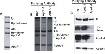
Trypanosome lytic factor (TLF) is a high-density lipoprotein (HDL) subclass providing innate protection to humans against infection by the protozoan parasite Trypanosoma brucei brucei. Two primate-specific plasma proteins, haptoglobin-related protein (Hpr) and apolipoprotein L-1 (ApoL-1), have been proposed to kill T. b. brucei both singularly or when co-assembled into the same HDL. To better understand the mechanism of T. b. brucei killing by TLF, the protein composition of TLF was investigated using a gentle immunoaffinity purification technique that avoids the loss of weakly associated proteins. HDL particles recovered by immunoaffinity absorption, with either anti-Hpr or anti-ApoL-1, were identical in protein composition and specific activity for T. b. brucei killing. Here, we show that TLF-bound Hpr strongly binds Hb and that addition of Hb stimulates TLF killing of T. b. brucei by increasing the affinity of TLF for its receptor, and by inducing Fenton chemistry within the trypanosome lysosome. These findings suggest that TLF in uninfected humans may be inactive against T. b. brucei prior to initiation of infection. We propose that infection of humans by T. b. brucei causes hemolysis that triggers the activation of TLF by the formation of Hpr-Hb complexes, leading to enhanced binding, trypanolytic activity, and clearance of parasites.


Conflict of interest statement

Competing interests. The authors have declared that no competing interests exist.

Figures

Figure 1
Figure 1. Analysis of Differentially Purified Human Serum HDLs
(A) TLF analyzed by non-reducing 10% SDS-PAGE and Coomassie brilliant blue staining after purification by a two-step purification process that includes high-salt density centrifugation and antibody affinity chromatography with anti-Hpr. The presence of albumin (Alb) and immunoglobulin (Ig) are indicated. (B) HDL purified from serum using a single-step affinity purification method with antibodies to ApoL-1, Hpr, or ApoA-1 and analyzed by non-reducing 10% SDS-PAGE and stained with Coomassie brilliant blue. Tf, transferrin. (C) The same gel as in (B) transferred to nitrocellulose and probed with antibodies to transferrin (Tf), Hpr, ApoL-1, and ApoA-1.
Figure 2
Figure 2. Lysis and Morphology of T. b. brucei Killed with Purified HDL
(A) Human HDLs were purified by a single-step immunoaffinity method with either anti-Hpr, anti-ApoL-1, or anti-ApoA-1. The specific activity for T. b. brucei killing was similar for TLF purified with the single-step protocol with either anti-Hpr (closed circles) or anti-ApoL-1 (open circles) (∼0.5 μg/ml killed 40% of the trypanosomes in our standard assay). The specific activity of samples purified with single-step immunoabsorption with anti-ApoA-1 (closed squares) was over 10-fold reduced in specific activity for T. b. brucei killing. (B) TLF purified by a two-step method using both high-salt density gradient ultracentrifugation and immunoaffinity absorption. TLF first subjected to high-salt gradient ultracentrifugation was 10-fold more toxic to T. b. brucei than TLF purified by the single-step immunoabsorption directly from serum. Similar specific activities were obtained when the second immunoaffinity step was conducted with either anti-Hpr (closed circles) or anti-ApoL-1 (open circles). The specific activity of HDLs purified with anti-ApoA-1 (closed squares) was over 100-fold lower. (C–F) Cell morphology of T. b. brucei treated with TLF purified by single-step immunoaffinity with anti-ApoL-1 or anti-Hpr. Following incubation at 37 °C for 2 h, cells were either smeared on microscope slides and methanol fixed (C and E) or directly viewed following imbedding in 1% agarose (D and F). Incubation with TLF purified either by anti-Hpr (C and D) or anti-ApoL-1 (E and F) immunoabsorption showed similar morphologies. Prior to lysis all cells progressed through a series of morphological changes that included transition to a kite-shaped form and continued swelling until lysis. The position of the kinetoplast (k) and nucleus (n) was determined by DAPI staining in the fixed preparations (C and E). The nucleus (n) was clearly visible in the unfixed cells (D and F). doi:110.1371/journal.ppat.0030129.g002
Figure 3
Figure 3. Binding of Purified Hpr and TLF to Hb
(A) SPR analysis of the binding of purified Hpr (left panel) and TLF purified by the two-step procedure (right panel) to immobilized Hb A0. The concentration of Hpr and TLF is shown to the right of the binding curves. The BIAevaluation 4.1 software estimated a Kd of 2–5 nM for the Hpr–Hb complex. Arrows indicate the beginning of the dissociation curves. (B) Silver-stained SDS-polyacrylamide (8%–16% gradient) gel of proteins eluted from Hb-coupled Sepharose (Hb), BSA-coupled Sepharose (BSA), or underivatized Sepharose (blank) after incubation with purified TLF. doi:110.1371/journal.ppat.0030129.g003
Figure 4
Figure 4. Effect of FBS on TLF Killing of T.b. brucei
(A) FBS (1 μl), BSA (1 μg), and Hb (1.0 μg) were fractionated on 15% SDS-PAGE gels and stained with Coomassie. The presence of Hb in FBS was verified by mass spectroscopy of the appropriate sized bands. Both Bos aurus alpha and gamma subunits of Hb were identified, and five of the gamma globin tryptic fragments are shown. (B) FBS is required for maximum in vitro lysis of T. b. brucei with highly purified TLF. FBS was titrated into a 300-μl assay containing 1% BSA. Addition of 5 μl FBS restored lytic levels to those seen in standard lysis assays [8]. (C) The effect of FBS addition is concentration dependent. TLF used in these studies was purified by the two-step procedure.
Figure 5
Figure 5. Hb Is the Co-Factor in FBS Necessary for Maximal TLF Killing of T. b. brucei
(A) Titration of Hb into in vitro lysis reactions containing highly purified TLF. Samples contained TLF (1.5 units) in 10% FBS (open circles); TLF (1.5 units) in 1% BSA and 1% glucose (closed circles); Hp in 1% BSA and 1% glucose without TLF (closed squares); and no TLF (Hb only) (open squares). In the presence of TLF, Hb addition results in a concentration-dependent increase in T. b. brucei killing. No increase is seen when Hb is added to FBS-containing reactions because FBS contains approximately 0.5 μg/μl of Hb. (B) Addition of Hp inhibits Hb-dependent killing by TLF. Hp was titrated into reactions containing TLF (1.5 units), 1% BSA and 1% glucose, and 0.1 pmol of Hb (closed circles). Addition of Hp and Hb (0.1 pmol) to T. b. brucei in the absence of TLF had no effect on cell viability (open circles). TLF used in these studies was purified by the two-step procedure.
Figure 6
Figure 6. Hb Is Required for Binding and Uptake of TLF
(A) Alexa Fluor 488–labeled TLF was incubated with trypanosomes and increasing amounts of Hb. Binding was measured by flow cytometry and the relative mean fluorescence intensity determined following binding for 1 h at 4 °C. In the absence of Hp, there is a dose-dependent increase of binding of Alexa Fluor 488–labeled TLF in the presence of Hb (closed circles). Addition of Hp inhibited TLF–Hb binding to T. b. brucei (open circles). (B) Uptake of TLF–Hb by T. b. brucei. Trypanosomes were pretreated with chloroquine for 30 min to block acidification of the lysosome and activation of TLF, followed by the addition of Alexa Fluor 594–labeled Hb and Alexa Fluor 488–labeled TLF. Cells were incubated for an additional hour at 37 °C. The TLF–Hb complex was endocytosed and traffics to the lysosome (Figure S2B). TLF used in these studies was purified by the two-step procedure.
Figure 7
Figure 7. TLF–Hb Induces Iron-Dependent Lysosomal Membrane Breakdown
(A) T. b. brucei was treated with increasing concentration of the iron chelator deferiprone in the presence (closed circles) and absence (open circles) of TLF–Hb. The inhibition of trypanosome lysis by deferiprone is indicated. We normalized the data to indicate that the addition of no deferiprone gave zero inhibition of TLF–Hb lysis after 2 h at 37 °C. (B) Trypanosomes were treated with increasing concentrations of the antioxidant DPPD in the presence (closed circles) or absence (open circles) of TLF–Hb. The inhibition of trypanosome lysis by DPPD is indicated. We normalized the data to indicate that the addition of no DPPD gave zero inhibition of TLF–Hb lysis after 2 h at 37 °C. (C) Effect of human serum and TLF–Hb treatment on T. b. brucei lysosomal membrane structure. In order to label the lysosome with a size-selective membrane-permeable marker, T. b. brucei was pre-incubated for 30 min at 37 °C with fluorescein-conjugated dextrans (500 kDa). Normal human serum (NHS) or purified TLF–Hb was then added. After 30 or 120 min, cells were formaldehyde fixed and stained with anti-p67 to localize the lysosomal membrane. After treatment with normal human serum or TLF–Hb for 30 min, dextrans are localized to a single cytoplasmic structure that stained with an antibody against the lysosomal marker p67. Trypanosomes treated for 120 min with either normal human serum or TLF–Hb continued to show p67 staining largely restricted to a single structure in the cell, but the 500-kDa dextrans are widely distributed in the cells, indicating a breakdown in lysosomal membranes. TLF used in these studies was purified by the two-step procedure.

Similar articles

Cited by

References

    1. Hoare CA. The Trypanosomes of mammals. A zoological monograph. Oxford: Blackwell Scientific Publications; 1972.
    1. Gibson W. Will the real Trypanosoma brucei rhodesiense please step forward? Trends Parasitol. 2002;18:486–490. - PubMed
    1. Gibson WC. Will the real Trypanosoma b. gambiense please stand up. Parasitol Today. 1986;2:255–257. - PubMed
    1. Pays E, Vanhamme L, Perez-Morga D. Antigenic variation in Trypanosoma brucei: Facts, challenges and mysteries. Curr Opin Microbiol. 2004;7:369–374. - PubMed
    1. Hajduk SL, Hager K, Esko JD. High-density lipoprotein-mediated lysis of trypanosomes. Parasitol Today. 1992;8:95–98. - PubMed

Publication types

MeSH terms