. 2007 Oct;101(4):262-8.
doi: 10.1111/j.1742-7843.2007.00116.x.

Silymarin prevents palmitate-induced lipotoxicity in HepG2 cells: involvement of maintenance of Akt kinase activation

Affiliations

Silymarin prevents palmitate-induced lipotoxicity in HepG2 cells: involvement of maintenance of Akt kinase activation

Zhenyuan Song et al. Basic Clin Pharmacol Toxicol. 2007 Oct.

Abstract

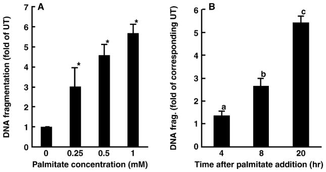
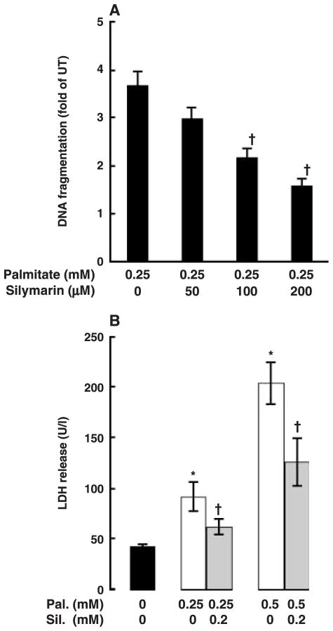
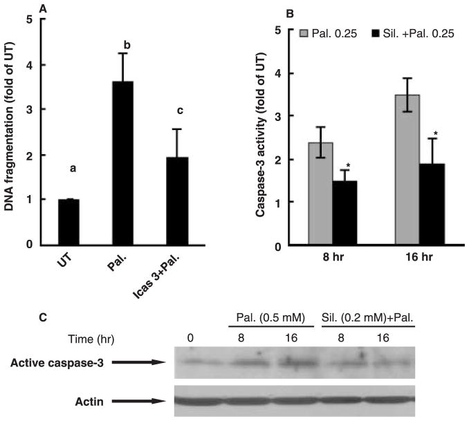
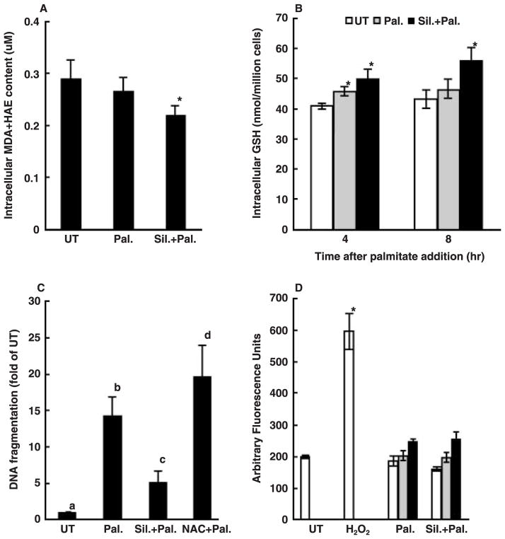
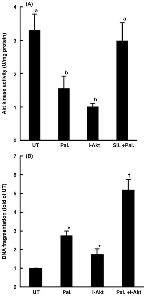
Whereas adipocytes have a unique capacity to store excess free fatty acids in the form of triglyceride in lipid droplets, non-adipose tissues, such as liver, have a limited capacity for storage of lipids. Saturated long-chain fatty acids, such as palmitate, are the major contributors to lipotoxicity. Silymarin is a mixture of flavonolignans, extracted from the milk thistle (Silibum marianum). Its hepatoprotective properties have been studied both in vitro and in vivo; however, its effect on palmitate-induced lipotoxicity has not been investigated. The objective of this study was to investigate (i) whether silymarin could protect HepG2 cells from palmitate-induced cell death in an in vitro model, and (ii) possible mechanisms involved in this hepatoprotective role of silymarin. HepG2 cells were treated with palmitate in the absence or presence of silymarin and supernatants or cell lysates were collected at varying time-points. Cell death was assayed by measuring DNA fragmentation, caspase-3 activity and lactate dehydrogenase release. Lipid peroxidation was assessed by measuring malondialdehyde and 4-hydroxyalkenals. Akt kinase activity was also measured. Incubation with palmitate caused significant death in HepG2 cells. Palmitate incubation did not cause significant changes in reactive oxygen species production or intracellular glutathione content, but markedly inhibited Akt kinase activity. Pre-treatment of HepG2 cells with silymarin prevented palmitate-induced inhibition of Akt kinase activity and attenuated cell death. Our results suggest that silymarin may be an effective agent in protecting hepatocytes from saturated fatty acids-induced cell death. These data also provide a further rationale for exploration of the use of silymarin in the treatment of non-alcoholic steatohepatitis.


Figures

Fig. 1
Fig. 1
Palmitate induces cell death in HepG2 cells. Cell death was measured by DNA fragmentation ELISA assay and expressed as fold of untreated. (A) Dose response of cell death induced by palmitate. HepG2 cells were incubated with increased concentration of palmitate (0, 0.25, 0.5 and 1 mM) for 20 hr. At least three experiments were conducted for each measurement. *Value is significantly different from that seen in control cells, P < 0.05. (B) Time-course change of cell death induced by palmitate. HepG2 cells were treated with 0.25 mM palmitate for different period. Values in bars that do not share a letter differ significantly. At least three experiments were conducted for each measurement.
Fig. 2
Fig. 2
Silymarin attenuates palmitate-induced cell death in HepG2 cells. (A) Silymarin protected HepG2 cells from palmitate-induced cell death in a dose-dependent manner. HepG2 cells were pre-treated with different dose of silymarin for 2 hr followed by 0.25 mM palmitate treatment for 20 hr. At least three experiments were conducted for each measurement. *Value is significantly different from that seen in control cells, P < 0.05. (B) Silymarin protects HepG2 cells from palmitate-induced lactate dehydrogenase (LDH) (U/l) release in a dose-dependent manner. HepG2 cells were pre-treated with a different dose of silymarin for 2 hr followed by 0.25 mM palmitate treatment for 20 hr. At least three experiments were conducted for each measurement. *Value is significantly different from that seen in control cells; Value is statistically different from that seen in the absence of silymarin, P < 0.05.
Fig. 3
Fig. 3
Effects of silymarin on palmitate-induced caspase-3 activation in HepG2 cell. (A) Caspase-3 inhibitor inhibited palmitate-induced cell death in HepG2. Apoptosis was measured by DNA fragmentation ELISA assay and expressed as fold of untreated. At least three experiments were conducted for each measurement. Values in bars that do not share a letter differ significantly, P < 0.05. Both (B) enzymatic activity assay and (C) Western blot analysis indicated that silymarin pre-treatments prevented palmitate-induced caspase-3 activation. *Value is statistically different from that seen in the absence of silymarin, P < 0.05.
Fig. 4
Fig. 4
Protective effect of silymarin on palmitate-induced cell death is independent of its antioxidant property. (A) Changes of intracellular malondialdehyde (MDA) + 4-hydroxyalkenals (HAE) content (μM). HepG2 cells were incubated with 0.25 mM palmitate for 20 hr with or without silymarin or N-acetylcysteine (NAC) pre-treatment. (B) Changes of intracellular glutathione (GSH) content (nmol/million cells). HepG2 cells were incubated with 0.25 mM palmitate for 4 and 8 hr, respectively, with or without silymarin or N-acetylcysteine (NAC) pre-treatment. *Value is significantly different from that seen in the presence of palmitate alone, P < 0.05. (C) NAC did not protect palmitate-induced cell death in HepG2 cells. Apoptosis was measured by DNA fragmentation ELISA assay and expressed as fold of untreated. Values in bars that do not share a letter differ significantly. (D) Changes of 2′,7′-dichlorohydrofluorescein diacetate fluorescent products. HepG2 cells were incubated with 300 μM H2O2 for 1 hr or with palmitate for 1, 4 and 20 hr, respectively. Data are expressed as Arbitrary Fluorescence Units. *Value is significantly different from that seen in the control cells, P < 0.05. At least three experiments were conducted for each measurement.
Fig. 5
Fig. 5
Maintenance of Akt kinase activation involved in the protective effect of silymarin on palmitate-induced cell death. (A) Silymarin prevented palmitate-induced inhibition on Akt kinase activation in HepG2 cells. Akt kinase activity (U/mg protein) was measured when incubation of HepG2 cells with 0.25 mM palmitate with or without silymarin pre-treatment. At least three experiments were conducted for each measurement. Values in bars that do not share a letter differ significantly, P < 0.05. (B) Inhibition of Akt kinase induced cell death in HepG2. At least three experiments were conducted for each measurement. *Value is significantly different from that seen in the control cells; Value is significantly different from that seen in the presence of palmitate alone, P < 0.05.

References

    1. Harrison SA, Neuschwander-Tetri BA. Nonalcoholic fatty liver disease and nonalcoholic steatohepatitis. Clin Liver Dis. 2004;8:861–79. - PubMed
    1. Dixon JB, Bhathal PS, O’Brien PE. Nonalcoholic fatty liver disease: predictors of nonalcoholic steatohepatitis and liver fibrosis in the severely obese. Gastroenterology. 2001;121:91–100. - PubMed
    1. Marchesini G, Brizi M, Morselli-Labate AM, et al. Association of nonalcoholic fatty liver disease with insulin resistance. Am J Med. 1999;107:450–5. - PubMed
    1. Choudhury J, Sanyal AJ. Insulin resistance in NASH. Front Biosci. 2005;10:1520–33. - PubMed
    1. Choudhury J, Sanyal AJ. Insulin resistance and the pathogenesis of nonalcoholic fatty liver disease. Clin Liver Dis. 2004;8:575–94. - PubMed

Publication types

MeSH terms