

doi: 10.1186/1742-4690-4-63.
Centrosomal pre-integration latency of HIV-1 in quiescent cells
Affiliations
- PMID: 17845727
- PMCID: PMC2014762
- DOI: 10.1186/1742-4690-4-63
Item in Clipboard
Centrosomal pre-integration latency of HIV-1 in quiescent cells
Retrovirology.
.
Abstract
Human immunodeficiency virus type 1 (HIV-1) efficiently replicates in dividing and non-dividing cells. However, HIV-1 infection is blocked at an early post-entry step in quiescent CD4+ T cells in vitro. The molecular basis of this restriction is still poorly understood. Here, we show that in quiescent cells, incoming HIV-1 sub-viral complexes concentrate and stably reside at the centrosome for several weeks. Upon cell activation, viral replication resumes leading to viral gene expression. Thus, HIV-1 can persist in quiescent cells as a stable, centrosome-associated, pre-integration intermediate.
Figures
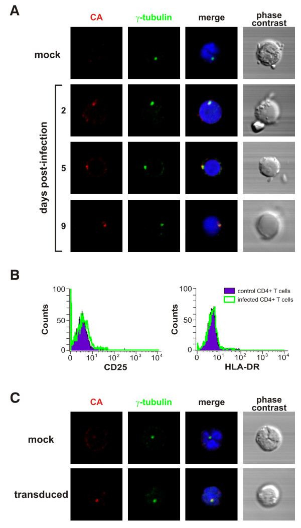
Sub-cellular localization of incoming HIV-1 in quiescent CD4+ T cells. A. Incoming HIV-1 CA localizes at the centrosome in infected human primary quiescent CD4+ T cells. Quiescent CD4+ T cells (0.5 × 10⁶ cells) were spinoculated with the NL4.3 strain of HIV-1 (moi = 1) as described [34]. The NL4.3 viral stock was obtained from 24-h harvests of supernatant from 293T cells transduced with a plasmid encoding the full-length viral genome and was titrated by limiting dilution MAGI assay [35]. At the indicated time points, infected and control cells were fixed in 4% PFA (15 min, 4°C), permeabilized with ice-cold methanol (5 min, 4°C) and stained with antibodies against HIV-1 CA protein (A25, Hybridolabs, Pasteur) and γ-tubulin (Abcam), a marker for the centrosome. Nuclei were stained with DAPI and images were acquired on a laser-scanning confocal microscope (LSM510 Meta; Carl Zeiss) equipped with an Axiovert 200 M inverted microscope, using a Plan Apo 63/1.4-N oil immersion objective. Co-localization between CA and γ-tubulin staining was observed in 58% to 75% of CA-positive cells. B) HIV-1 infection did not significantly alter the activation status of quiescent CD4+ T cells. Surface expression of T cell activation markers (CD25 and HLA-DR) was monitored by flow cytometry. C) Pericentriolar distribution of incoming HIV-1 CA in quiescent CD4+ T cells transduced with a VSVg-pseudotyped HIV-1 based lentivector carrying the GFP transgene. The lentivector stock was produced by co-transfected with an HIV-derived packaging construct, the VSVg-expressor vector and the plasmid vector (psPAX2, pMD2.G and pWPI, respectively, a gift from D. Trono), as described [35]. The titre of the lentivector stocks was determined by measuring the percentage of GFP positive cells 48 h following transduction of 293T cells by flow cytometry. Transduced and control quiescent CD4+ T cells were immunostained and visualized as described above. Co-localization between CA and γ-tubulin staining was observed in 60% to 82% of CA-positive cells.
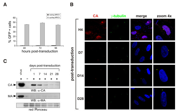
Incoming HIV-1 persistently reside at the centrosome of resting cells. A. Cycling but not resting MRC5 cells support viral gene expression. To obtain a resting cell population, MRC5 were grown to confluence, growth-arrested by serum starvation and cultured in the presence of 10-6 M dexamethasone. MRC5 cells were transduced with a VSVg-pseudotyped lentiviral vector carrying the GFP reporter gene and GFP-expression was measured by flow cytometry at 48, 72 and 96 h post-transduction. B. Incoming HIV-1 CA localizes at the centrosome in transduced MRC5 cells. Cells were immunostained and analyzed by confocal microscopy as described above. C. HIV-1 CA but not MA protein can be detected in the total cell extracts of transduced resting MRC5 up to 28 days post-transduction. Total cell extracts were obtained by boiling both transduced and control cells, pre-treated with pronase (10 min, 4°C), in SDS-PAGE sample buffer. Proteins were resolved by SDS-PAGE and detected by Western blotting with mouse anti-MA or mouse anti-CA ab.
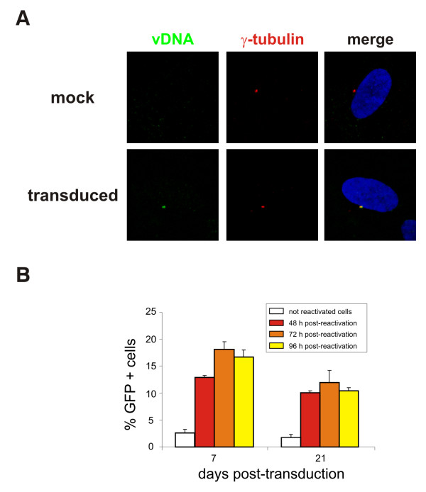
Centrosome-associated HIV-1 pre-integration intermediate is inducible upon cell activation. A. HIV-1 reverse-transcribed viral cDNA localizes at the centrosome of resting MRC5 cells transduced with a DNAse-treated VSVg-pseudotyped NL4.3 virus, which was made using the NL4.3Luc plasmid, in which the env gene was replaced by the luciferase transgene, and a VSVg-expressor vector. Fluorescence in situ hybridization (FISH) was performed 4 days after transduction using the full-length proviral genome as a probe [32]. After FISH, immunostaining with anti-γ-tubulin ab was performed as described above. B. Viral gene expression resumes after reactivation of quiescent cells. Transduced resting MRC5 cells were sorted to recover only GFP-negative cells which were then stimulated to divide by splitting and serum addition. The percentage of GFP-expressing cells was determined at 48, 72 and 96 h after sorting and reactivation by flow cytometry.
References
Publication types
MeSH terms
LinkOut - more resources
Full Text Sources
Research Materials
