. 2008 Feb;22(1):18-27.
doi: 10.1016/j.tiv.2007.07.004. Epub 2007 Jul 21.

Manganese-induced potentiation of in vitro proinflammatory cytokine production by activated microglial cells is associated with persistent activation of p38 MAPK

Affiliations

Manganese-induced potentiation of in vitro proinflammatory cytokine production by activated microglial cells is associated with persistent activation of p38 MAPK

P L Crittenden et al. Toxicol In Vitro. 2008 Feb.

Abstract

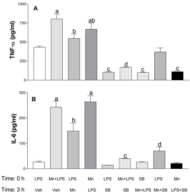
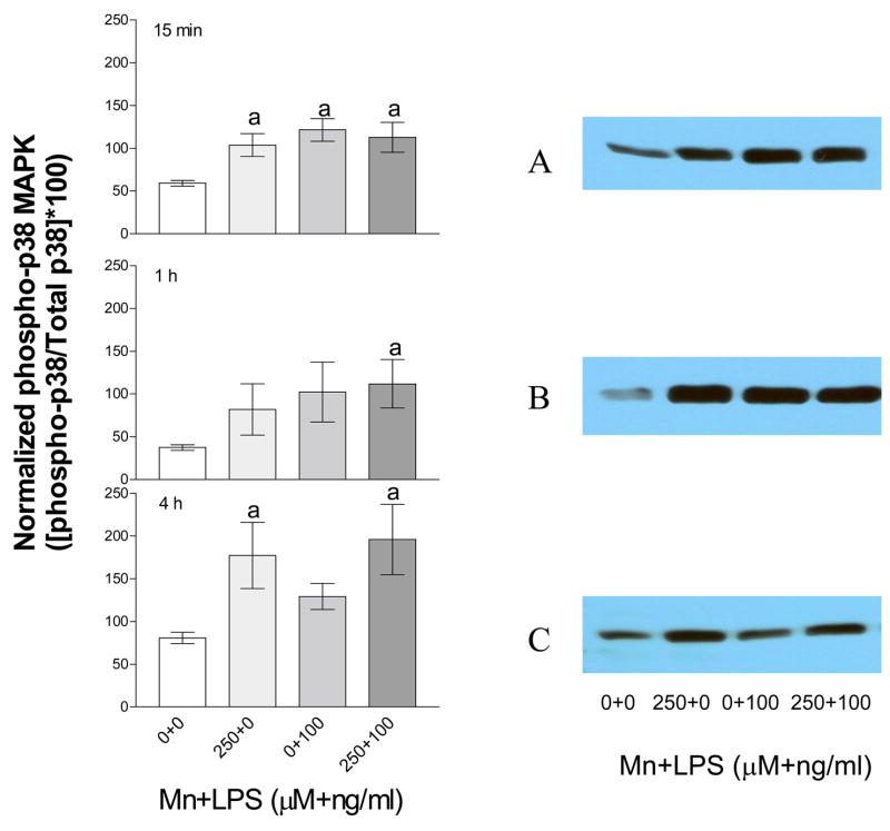
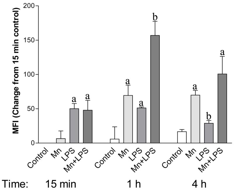
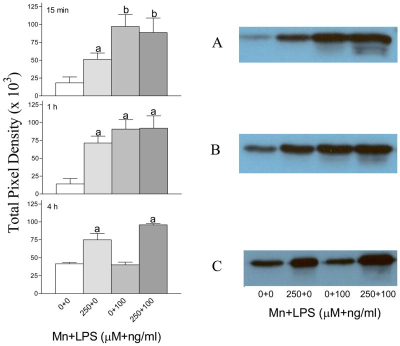
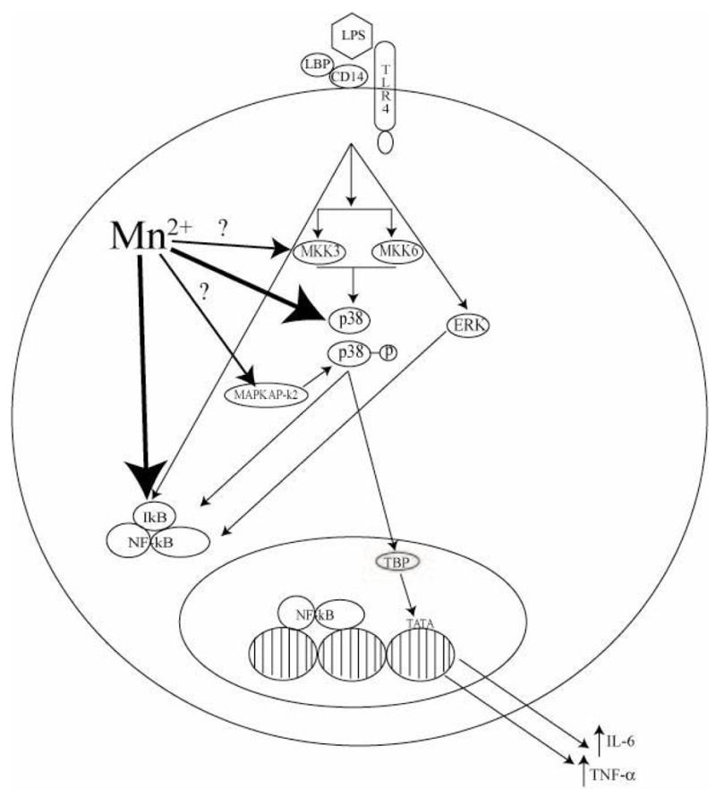
Previous studies that investigated the role of inflammation in the neurotoxicity of manganese (Mn) found that Mn enhanced the production of inflammogen (lipopolysaccharide; LPS)-induced proinflammatory cytokines such as IL-6 and TNF-alpha. Although we have shown that the enhanced cytokine production occurs via a NF-kappaB-dependent mechanism, the role of upstream kinases in this Mn-induced enhancement has not been explored. As other studies have demonstrated that p38 mitogen activated protein kinase (p38) is necessary for LPS-induced, NF-kappaB-dependent expression of proinflammatory cytokines, we hypothesized that Mn enhancement of LPS-induced production of IL-6 and TNF-alpha may be associated with p38 activation and conducted a series of experiments to address our hypothesis. We found that pre-treatment of microglial cells with a p38-inhibitor (SB203580) prevented Mn+LPS-induced production of IL-6 and TNF-alpha. Moreover, potentiation of IL-6 and TNF-alpha production, which occurred in both concurrent and sequential (3h apart) exposures to Mn and LPS, was inhibited by inhibition of p38. Additionally, Mn exposure enhanced the phosphorylation and activity of p38 and this effect was persistent. Although p38 activity declined over time LPS-exposed cells, it persisted in cells exposed to Mn or Mn+LPS. Thus, the increased production of proinflammatory cytokines by LPS-activated microglia exposed to Mn is associated with increased and persistent activation of p38.


Figures

Figure 1
Figure 1
Effect of delayed exposure to manganese (Mn) and/or SB203580 (SB, a p38 inhibitor) on lipopolysaccharide (LPS) activated microglial cytokine production. TNF-α (1A) and IL-6 (1B) production was analyzed in supernatants collected 24 h after exposure of N9 microglial cells to LPS (100 ng/ml, time 0) then vehicle (Veh, 3 h later), Mn (250 μM) + LPS (time 0) then Veh (3 h later), LPS (time 0) then Mn (3 h later), Mn (time 0) then LPS (3 h later), LPS (time 0) then SB (50 μM, 3 h later), LPS (time 0) then Mn + SB (3 h later), Mn (time 0) then LPS + SB (3 h later), Mn + LPS (time 0) then SB (3 h later), and SB (time 0) then Mn + LPS (3 h later). Media levels of TNF-α and IL-6 were analyzed by respective DuoSet ELISA as described in the Materials and Methods. Data shown in each bar represent the mean ± S.E.M. of 4–8 independent replicates. a, b, c, d Presence of letters on top of bars indicate treatment differences in the TNF-α (1A) and IL-6 (1B) levels with bars with different letters being different from each other and from bars without a letter (P < 0.05).
Figure 2
Figure 2
Densitometric analysis (left panels) and representative western blots (right panels) of phosphorylated p38 kinase in N9 microglia exposed to Mn, LPS, or Mn+LPS. Phosphorylated p38 protein from N9 cells exposed to vehicle, 250 μM Mn, 100 ng/ml LPS, or 250 μM Mn+100 ng/ml LPS for 15 min (A), 1 h (B) and 4 h (C) was determined by western blot analysis. Western blots and densitometric analyses were performed as described in the Materials and Methods. Data shown in each bar represent the mean ± S.E.M. of the adjusted pixel density for phosphorylated p38 normalized to non-phosphorylated p38. For abbreviations please refer to the legend on Figure 1. Each bar represents a minimum of 3 independent replicates. a Presence of letters on top of bars indicate treatment differences in phosphorylated p38 levels within a time point with bars with different letters being different from each other and from bars without a letter (P < 0.05).
Figure 3
Figure 3
Phosphorylated p38 protein levels in whole cell lysate of N9 microglia exposed to 250 μM with or without 100 ng/ml LPS. Whole cell lysates collected after 15 min, 1 h, or 4 h were analyzed by flow cytometry as described in the Materials and Methods. Data shown represent the mean fluorescence intensity (MFI) change from the 15 min control ± S.E.M. for two independent experiments where each experimental condition was independently replicated (n = 4). For abbreviations please refer to the legend on Figure 1. a, b Presence of letters on top of bars indicate treatment differences in phosphorylated p38 levels within a time point with bars with different letters being different from each other and from bars without a letter (P < 0.05).
Figure 4
Figure 4
p38 activity following exposure to vehicle, 250 μM Mn and/or 100 ng/ml LPS for 15 min (A), 1 h (B), or 4 h (C) in N9 microglial cells. The p38 activity in whole cell lysates was analyzed by western blot for the detection of a phosphorylated p38 substrate (ATF-2 Fusion Protein) followed by densitometric analyses (left hand panel) as described in the Materials and Methods. Blots representative of three independent experiments are shown in the right hand panel. Data shown in each bar represent the mean ± S.E.M. of the total pixel density for 3 independent replicates. For abbreviations please refer to the legend on Figure 1. a,b Presence of letters on top of bars indicate treatment differences in p38 activity within a time point with bars with different letters being different from each other and from bars without a letter (P < 0.05).
Figure 5
Figure 5
Diagram of potential/known Mn sites of action in microglial intracellular signaling following activation with LPS. Small arrows indicate known intracellular signaling pathways while larger arrows indicate possible sites of interaction between signaling molecules and Mn. Large arrows with a ‘?’ denote potential sites of action for Mn-enhanced p38 acitivity. Abbreviations: LPS (Lipopolysaccharide), LBP (LPS Binding Protein), TLR4 (Toll Like Receptor 4), MKK3/6 (Mitogen Activated Protein Kinase Kinase) p38 (p38 Mitogen Activated Protein Kinase), p38-p (phosphorylated p38), ERK (Extracellular Signal-Regulated Kinase), MAPKAP-k2 (MAPK Phosphatase), IkB (Inhibitor of NF-kB), NF-kB (Nuclear Factor kappa B), TBP (TATA-Binding Protein), TATA (TATA Box).

Similar articles

Cited by

References

    1. Aschner M. Manganese: Brain transport and emerging research needs. Environmental Health Prespectives. 2000;108(Suppl 3):429–432. - PMC - PubMed
    1. Aschner M, Aschner JL. Manganese neurotoxicity: Cellular effects and blood-brain barrier transport. Neuroscience Biobehavior Review. 1991;15:334–340. - PubMed
    1. Bae JH, Jang BC, Suh SI, Ha E, Baik HH, Kim SS, Lee MY, Shin DH. Manganese induces inducible nitric oxide synthase (iNOS) expression via activation of both MAP kinase and PI3K/Akt pathways in BV2 microglial cells. Neuroscience Letters. 2006;398:151–154. - PubMed
    1. Bhat NR, Zhang P, Lee JC, Hogan EL. Extracellular signal-regulated kinase and p38 subgroups of mitogen-activated protein kinases regulate inducible nitric oxide synthase and tumor necrosis factor-a gene expression in endotoxin-stimulated primary glial cultures. Journal of Neuroscience. 1998;18:1633–1641. - PMC - PubMed
    1. Carter AB, Knudtson KL, Monick MM, Hunninghake GW. The p38 mitogen-activated protein kinase is required for NF-κB-dependent gene expression. Journal of Biological Chemistry. 1999;274:30858–30863. - PubMed

Publication types

MeSH terms