

Hippocampal activation in adults with mild cognitive impairment predicts subsequent cognitive decline
- PMID: 17846109
- PMCID: PMC2683145
- DOI: 10.1136/jnnp.2007.124149
Hippocampal activation in adults with mild cognitive impairment predicts subsequent cognitive decline
Abstract
Objective: To use functional MRI (fMRI) to investigate whether hippocampal activation during a memory task can predict cognitive decline in individuals with mild cognitive impairment (MCI).
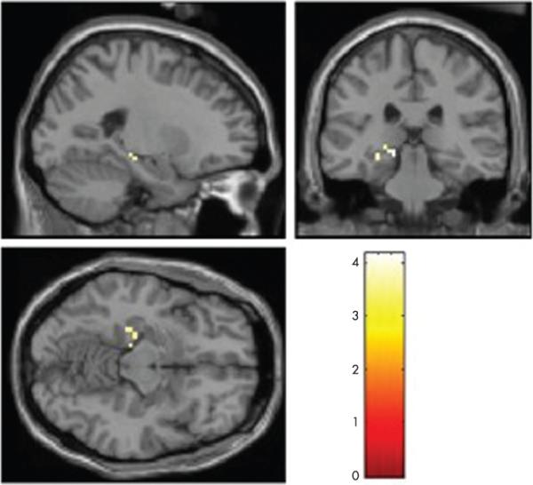
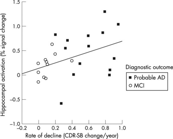
Methods: 25 older individuals with MCI performed a visual scene encoding task during fMRI scanning, and were followed clinically for at least 4 years after scanning. A hypothesis driven analysis of fMRI data was performed. First, fMRI data were analysed at the group level to identify the regions of the hippocampal formation that were engaged by this memory task. Parameter estimates of each subject's memory related hippocampal activation (% signal change) were extracted and were analysed with a linear regression model to determine whether hippocampal activation predicted the degree or rate of cognitive decline, as measured by change in Clinical Dementia Rating Sum-of-Boxes (CDR-SB).
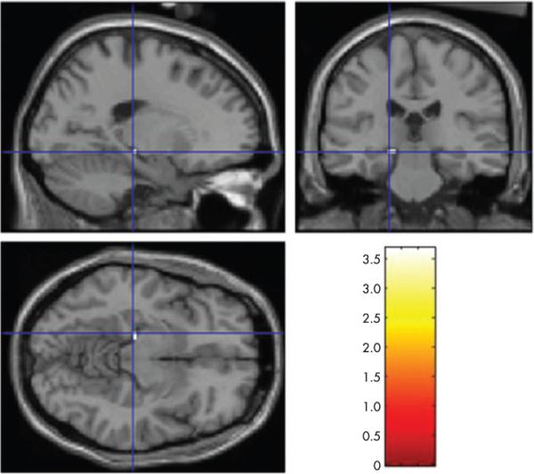
Results: Over 5.9 (1.2) years of follow-up after scanning, subjects varied widely in degree and rate of cognitive decline (change in CDR-SB ranged from 0 to 6, and the rate ranged from 0 to 1 CDR-SB unit/year). Greater hippocampal activation predicted greater degree and rate of subsequent cognitive decline (p<0.05). This finding was present even after controlling for baseline degree of impairment (CDR-SB), age, education and hippocampal volume, as well as gender and apolipoprotein E status. In addition, an exploratory whole brain analysis produced convergent results, demonstrating that the hippocampal formation was the only brain region where activation predicted cognitive decline.
Conclusions: In individuals with MCI, greater memory task related hippocampal activation is predictive of a greater degree and rate of cognitive decline subsequent to scanning. fMRI may provide a physiological imaging biomarker useful for identifying the subgroup of MCI individuals at highest risk of cognitive decline for potential inclusion in disease modifying clinical trials.
Figures
References
-
- Dickerson BC, Goncharova I, Sullivan MP, et al. MRI-derived entorhinal and hippocampal atrophy in incipient and very mild Alzheimer's disease. Neurobiol Aging. 2001;22:747–54. - PubMed
-
- Killiany RJ, Gomez-Isla T, Moss M, et al. Use of structural magnetic resonance imaging to predict who will get Alzheimer's disease. Ann Neurol. 2000;47:430–9. - PubMed
-
- Visser PJ, Scheltens P, Verhey FR, et al. Medial temporal lobe atrophy and memory dysfunction as predictors for dementia in subjects with mild cognitive impairment. J Neurol. 1999;246:477–85. - PubMed
Publication types
MeSH terms
Grants and funding
LinkOut - more resources
Full Text Sources
Other Literature Sources
Medical