. 2007 Nov;75(11):5200-9.
doi: 10.1128/IAI.00954-07. Epub 2007 Sep 10.

Presentation of Toxoplasma gondii antigens via the endogenous major histocompatibility complex class I pathway in nonprofessional and professional antigen-presenting cells

Affiliations

Presentation of Toxoplasma gondii antigens via the endogenous major histocompatibility complex class I pathway in nonprofessional and professional antigen-presenting cells

Florence Dzierszinski et al. Infect Immun. 2007 Nov.

Abstract

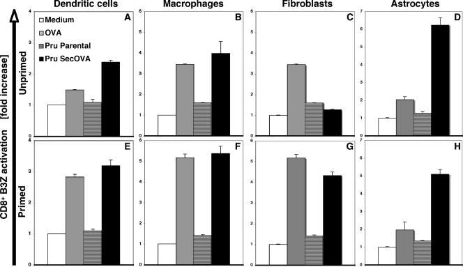
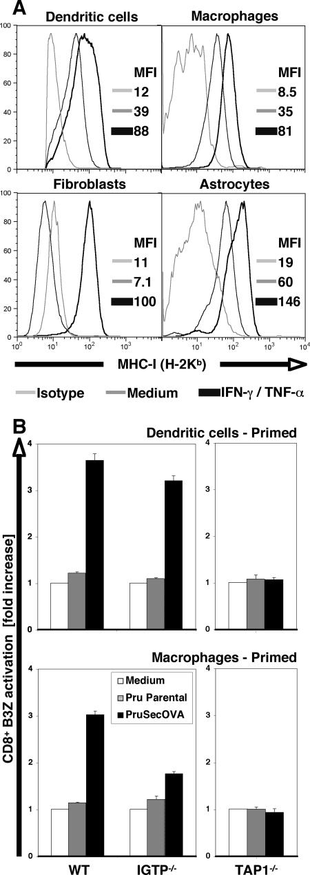
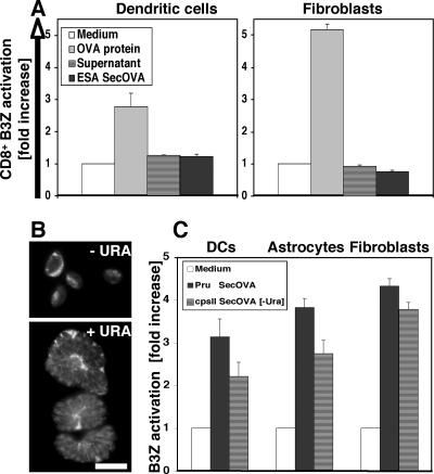
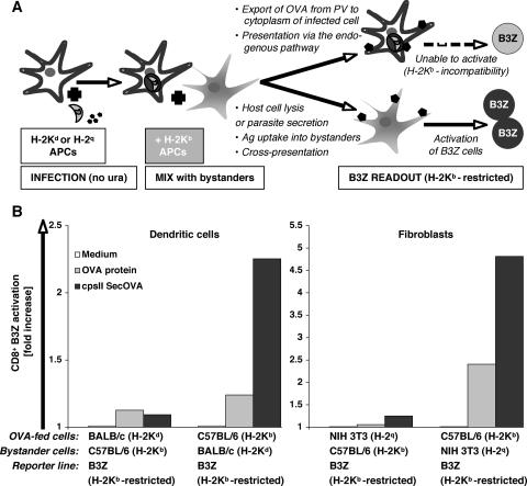
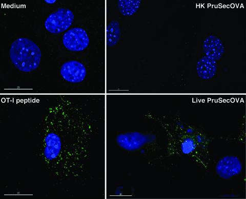
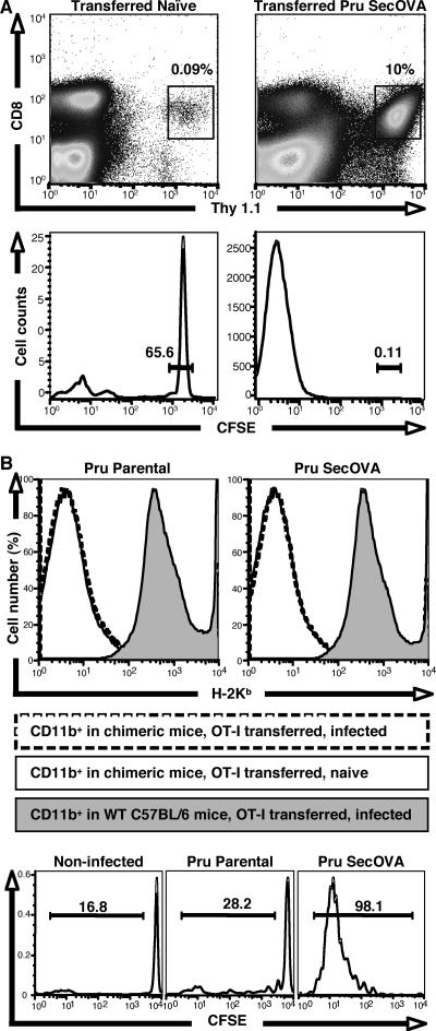
Challenge with the intracellular protozoan parasite Toxoplasma gondii induces a potent CD8+ T-cell response that is required for resistance to infection, but many questions remain about the factors that regulate the presentation of major histocompatibility complex class I (MHC-I)-restricted parasite antigens and about the role of professional and nonprofessional accessory cells. In order to address these issues, transgenic parasites expressing ovalbumin (OVA), reagents that track OVA/MHC-I presentation, and OVA-specific CD8+ T cells were exploited to compare the abilities of different infected cell types to stimulate CD8+ T cells and to define the factors that contribute to antigen processing. These studies reveal that a variety of infected cell types, including hematopoietic and nonhematopoietic cells, are capable of activating an OVA-specific CD8+ T-cell hybridoma, and that this phenomenon is dependent on the transporter associated with antigen processing and requires live T. gondii. Several experimental approaches indicate that T-cell activation is a consequence of direct presentation by infected host cells rather than cross-presentation. Surprisingly, nonprofessional antigen-presenting cells (APCs) were at least as efficient as dendritic cells at activating this MHC-I-restricted response. Studies to assess whether these cells are involved in initiation of the CD8+ T-cell response to T. gondii in vivo show that chimeric mice expressing MHC-I only in nonhematopoietic compartments are able to activate OVA-specific CD8+ T cells upon challenge. These findings associate nonprofessional APCs with the initial activation of CD8+ T cells during toxoplasmosis.


Figures

FIG. 1.
FIG. 1.
MHC-I presentation of OVA by hematopoietic and nonhematopoietic cells infected with OVA transgenic T. gondii. DCs, MΦ, fibroblasts, or astrocytes were cultivated without (top panels) or with (bottom panels) IFN-γ and TNF-α, followed by 16 h of incubation with 500 μg/ml OVA protein, 5 × 10⁴ Prugniaud ΔHXGPRT strain (Pru) T. gondii tachyzoites, or 5 × 10⁴ Prugniaud ΔHXGPRT strain T. gondii engineered to express SecOVA. Professional APCs were prepared from the BM of C57BL/6 mouse (H-2Kb) MHC-I, and OVA presentation was assayed using a B3Z CD8+ T-cell hybridoma that specifically expresses LacZ upon interaction with MHC-I (Kb)/OVA257-264 (SIINFEKL) complexes (25). Results (averages for at least five experiments with triplicate samples ± standard deviation [SD]) are expressed as the increase (n-fold) in the OD at 562 nm (OD562) relative to samples where antigen (but not B3Z cells) was omitted (white bars). In contrast to professional APCs, fibroblasts require priming in order to present SecOVA to B3Z cells. At the time when OD562 values are read for B3Z cell activation (48 h postinfection), parasites have replicated but have not lysed out from the originally infected APCs; if assays were left until host cell lysis and reinfection of other cells (including T cells) occurred, then the negative-control Prugniaud ΔHXGPRT parental strain samples (striped bars) would become positive (wells would turn from yellow to purple).
FIG. 2.
FIG. 2.
The relationship between IFN-γ and MHC-I presentation of SecOVA. (A) Cell surface expression of MHC-I (H-2Kb) molecules in unprimed or IFN-γ/TNF-α primed BM-DCs, BM-MΦ, fibroblasts, and astrocytes, determined by flow cytometry. MFI, mean fluorescence intensity. Note the strong induction in primed nonhematopoietic cells. This is consistent with the hypothesis that, in fibroblasts, priming (i) is required to activate SecOVA presentation via the endogenous MHC-I pathway and (ii) enhances the presentation of exogenous OVA via cross-presentation (Fig. 1C and 1G). In DCs, this activation of the endogenous MHC-I pathway is not required for SecOVA presentation, but it does enhance both endogenous and cross-presentation pathways (Fig. 1A and 1E). (B) BM-DCs (top) and BM-MΦ (bottom) from IGTP- or TAP1-deficient mice were primed with IFN-γ/TNF-α and assessed for the ability to present SecOVA to B3Z cells in vitro. Results are expressed as increases (n-fold) relative to results for controls incubated without antigen (white bars). WT, wild type.
FIG. 3.
FIG. 3.
MHC-I presentation of SecOVA from intracellular parasites versus lysed cell/parasite supernatant. (A) IFN-γ/TNF-α-primed H-2Kb BM-DCs and fibroblasts were incubated for 16 h with either OVA protein (500 μg/ml), concentrated supernatant from a lysed-out culture of ∼5 × 10⁷ Prugniaud ΔHXGPRT SecOVA parasites, or excreted/secreted antigens from 5 × 10⁴ Prugniaud ΔHXGPRT SecOVA tachyzoites, and MHC-I/OVA presentation was measured using the B3Z cell assay; the number of parasites used corresponds to conditions employed in antigen presentation assays. B3Z cells were activated only when APCs were incubated with large amounts of exogenous soluble OVA (shown are averages for three experiments performed in triplicate ± SD). (B) RHΔcpsII T. gondii parasites (15) engineered to secrete OVA into the PV were incubated with or without 0.2 mM uracil (URA) prior to staining with polyclonal anti-OVA. In these uracil auxotrophs, replication (and therefore cytolysis) is dependent on uracil, but parasites remain alive for several days, continuing to metabolize and secrete OVA protein into the PV. Bar = 10 μm. (C) IFN-γ/TNF-α-primed H-2Kb BM-DCs, astrocytes, and fibroblasts were incubated for 16 h with 5 × 10⁴ wild-type Prugniaud ΔHXGPRT (Pru) SecOVA parasites or 5 × 10⁴ nonreplicating RHΔcpsII SecOVA parasites in the absence of uracil (−Ura). MHC-I presentation of SecOVA (detected using the B3Z cell assay) did not require parasite replication or host cell cytolysis, suggesting an endogenous origin of the presented antigen (average results from five or more experiments performed in triplicate ± SD).
FIG. 4.
FIG. 4.
Actively infected cells, rather than bystanders, present T. gondii SecOVA in the context of MHC-I. (A) Experimental design for a haplotype-mixing experiment based on the restricted reactivity of B3Z CD8+ T cells to H-2Kb MHC-I molecules. First, 105 IFN-γ/TNF-α-primed APCs bearing haplotypes H-2Kd (BALB/c BM-DCs) or H-2q (NIH 3T3 fibroblasts) are incubated with OVA protein (500 μg/ml) or infected with RHΔcpsII SecOVA parasites (MOI = 0.5) and incubated for 12 h to permit invasion. These cells are then mixed with 105 APCs bearing the H-2Kb haplotype, which are capable of activating B3Z cells (reacting with the H-2Kb haplotype). In theory, lysis of the infected host cell or antigen secretion in the medium, followed by antigen uptake by bystander APCs, could activate B3Z cells via cross-presentation (lower pathway). Infected (or OVA-treated) cells are unable to activate B3Z cells by either the endogenous pathway or lysis and cross-presentation, due to H-2K incompatibility. (B) When APCs expressing H-2Kd (BALB/c BM-DC) or H-2q (NIH 3T3 cell) haplotypes were exposed to antigen, no cross-presentation was observed in vitro. If washes were omitted from the protocol before the addition of bystander cells, B3Z cell activation occurred, because H-2Kb APCs were then in direct contact with antigens or parasites (not shown). As the control, H-2Kb BM-DCs or fibroblasts were exposed to antigen (OVA peptide or SecOVA parasites), resulting in strong activation (average from three independent experiments). cpsII, RHΔcpsII.
FIG. 5.
FIG. 5.
MHC-I presentation of SecOVA only in host cells actively infected with T. gondii transgenics. Visualization of MHC-I/SIINFEKL complexes in primed astrocyte cultures that were untreated (top left), incubated with the SIINFEKL OT-I peptide (bottom left), exposed to heat-killed Prugniaud ΔHXGPRT SecOVA (PruSecOVA) parasites (top right), or actively infected with live Prugniaud ΔHXGPRT SecOVA parasites (bottom right). Green staining indicates MHC-I/SIINFEKL complexes detected using MAb 25D1.16 (43); blue indicates DAPI staining of host and parasite DNA. Note the intense staining of MHC-I/SIINFEKL complexes in cells that contain several PVs with replicating Prugniaud ΔHXGPRT SecOVA parasites. These clusters are still unidentified and would be MHC-I/SIINFEFL complexes either at the cell surface (concentrated in specific areas) and/or in the exocytic/endocytic pathways, because incubation with the 25D1.16 MAb was carried out using live cells, and MHC/peptide complexes may be internalized during this incubation. Uninfected (bystander) cells were not labeled.
FIG. 6.
FIG. 6.
Expansion and activation of CD8+ T cells in mice infected with T. gondii expressing SecOVA. (A) MHC-I/SIINFEKL-restricted OT-I Thy1.1 CD8+ T cells were labeled with CFSE and adoptively transferred to C57BL/6 Thy1.2 mice. Eight days after infection with Prugniaud ΔHXGPRT (Pru) SecOVA parasites (right panels) or mock treatment (left panels), cells in the draining mesenteric lymph nodes were stained for CD3, CD8, and Thy1.1 (top), and the positive populations (boxed) were assessed for retention of CFSE (bottom). Far more OVA-specific CD8+ T cells were observed in Prugniaud ΔHXGPRT SecOVA-infected animals than in naïve controls, and dilution of CFSE confirms proliferation of these cells. (B) Irradiated wild-type C57BL/6 mice were reconstituted with BM from β2-microglobulin-deficient animals, producing chimeras expressing MHC-I in nonhematopoietic tissues only. Chimeric mice were then infected with Prugniaud ΔHXGPRT parental or Prugniaud ΔHXGPRT SecOVA parasites 1 day after adoptive transfer of CFSE-labeled OT-I T cells. H-2Kb surface staining assayed by flow cytometry of the CD11b+ population from the mesenteric lymph nodes (top panels) shows minimal levels of MHC-I expression in the chimeric mice (no shading) relative to that seen with the wild-type C57BL/6 controls (shaded), regardless of infection status (solid versus broken lines). Nevertheless, CFSE dilution shows proliferation of the CD8+ OT-I T-cell population in the mesenteric lymph nodes 8 days after infection with Prugniaud ΔHXGPRT SecOVA (lower right), but not in uninfected (left) or Prugniaud ΔHXGPRT parental strain-infected animals (center).

References

    1. Arrode, G., C. Boccaccio, J. Lule, S. Allart, N. Moinard, J.-P. Abastado, A. Alam, and C. Davrinche. 2000. Incoming human cytomegalovirus pp65 (UL83) contained in apoptotic infected fibroblasts is cross-presented to CD8+ T cells by dendritic cells. J. Virol. 74:10018-10024. - PMC - PubMed
    1. Bertholet, S., R. Goldszmid, A. Morrot, A. Debrabant, F. Afrin, C. Collazo-Custodio, M. Houde, M. Desjardins, A. Sher, and D. Sacks. 2006. Leishmania antigens are presented to CD8+ T cells by a transporter associated with antigen processing-independent pathway in vitro and in vivo. J. Immunol. 177:3525-3533. - PubMed
    1. Bevan, M. J. 1976. Cross-priming for a secondary cytotoxic response to minor H antigens with H-2 congenic cells which do not cross-react in the cytotoxic assay. J Exp. Med. 143:1283-1288. - PMC - PubMed
    1. Boehm, U., T. Klamp, M. Groot, and J. C. Howard. 1997. Cellular responses to interferon-gamma. Annu. Rev. Immunol. 15:749-795. - PubMed
    1. Brown, C. R., C. A. Hunter, R. G. Estes, E. Beckmann, J. Forman, C. David, J. S. Remington, and R. McLeod. 1995. Definitive identification of a gene that confers resistance against Toxoplasma cyst burden and encephalitis. Immunology 85:419-428. - PMC - PubMed

Publication types