. 2007 Oct 1;204(10):2349-62.
doi: 10.1084/jem.20062596. Epub 2007 Sep 10.

Functionally specialized junctions between endothelial cells of lymphatic vessels

Affiliations

Functionally specialized junctions between endothelial cells of lymphatic vessels

Peter Baluk et al. J Exp Med. .

Abstract

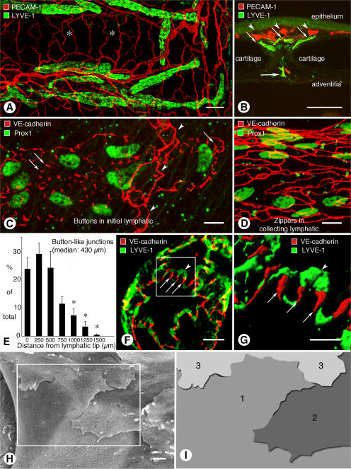
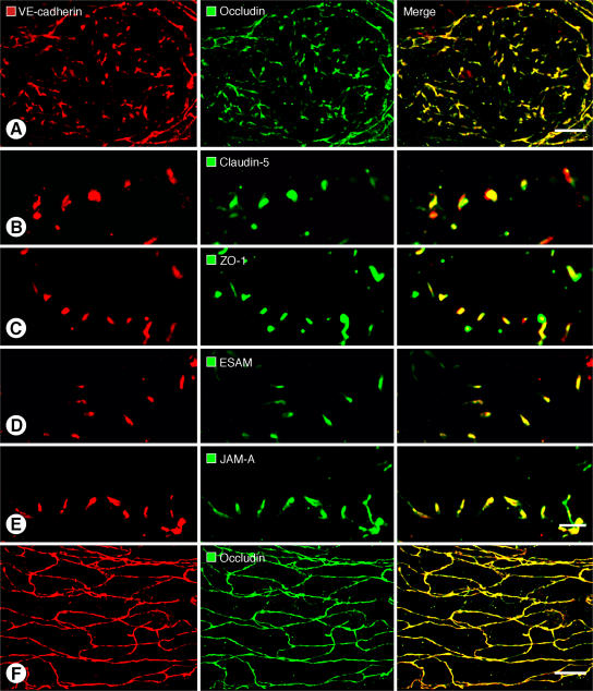
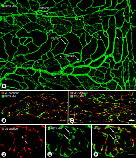
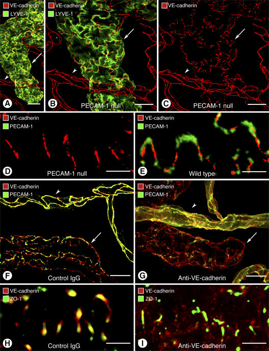
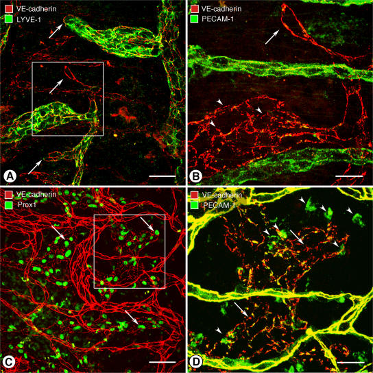
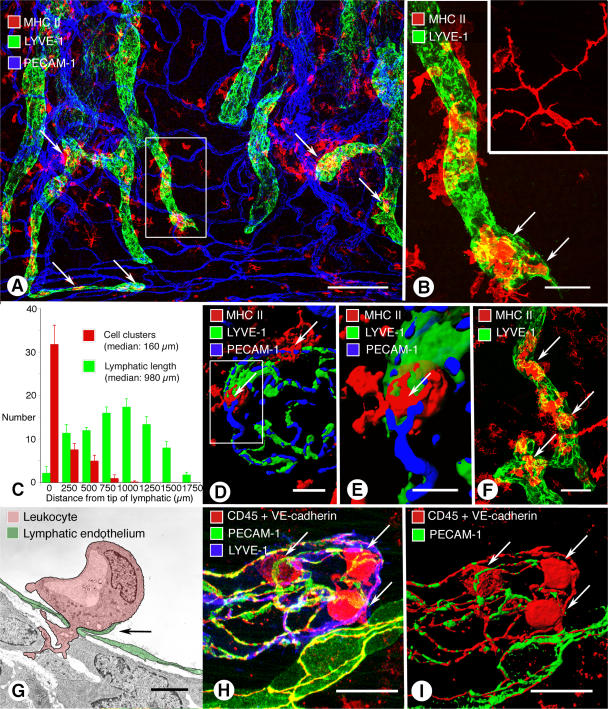
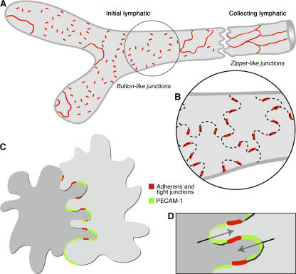
Recirculation of fluid and cells through lymphatic vessels plays a key role in normal tissue homeostasis, inflammatory diseases, and cancer. Despite recent advances in understanding lymphatic function (Alitalo, K., T. Tammela, and T.V. Petrova. 2005. Nature. 438:946-953), the cellular features responsible for entry of fluid and cells into lymphatics are incompletely understood. We report the presence of novel junctions between endothelial cells of initial lymphatics at likely sites of fluid entry. Overlapping flaps at borders of oak leaf-shaped endothelial cells of initial lymphatics lacked junctions at the tip but were anchored on the sides by discontinuous button-like junctions (buttons) that differed from conventional, continuous, zipper-like junctions (zippers) in collecting lymphatics and blood vessels. However, both buttons and zippers were composed of vascular endothelial cadherin (VE-cadherin) and tight junction-associated proteins, including occludin, claudin-5, zonula occludens-1, junctional adhesion molecule-A, and endothelial cell-selective adhesion molecule. In C57BL/6 mice, VE-cadherin was required for maintenance of junctional integrity, but platelet/endothelial cell adhesion molecule-1 was not. Growing tips of lymphatic sprouts had zippers, not buttons, suggesting that buttons are specialized junctions rather than immature ones. Our findings suggest that fluid enters throughout initial lymphatics via openings between buttons, which open and close without disrupting junctional integrity, but most leukocytes enter the proximal half of initial lymphatics.


Figures

Figure 1.
Figure 1.
Buttons in endothelium of initial lymphatics. (A) Confocal images showing lymphatic vessels (green, LYVE-1) and blood vessels (red, PECAM-1) in whole mount of mouse trachea. Region of mucosa over horizontal cartilage (*) is mostly free of lymphatics. (B) Longitudinal section of trachea shows epithelium (green), subepithelial blood vessels (red, arrowheads), more deeply positioned initial lymphatics (diagonal arrows), collecting lymphatic (horizontal arrow), and adjacent cartilages. (C and D) Confocal images of VE-cadherin immunoreactivity (red) at discontinuous buttons in initial lymphatic (arrows; C) and continuous zippers in collecting lymphatic (D). Zippers are also present in blood capillary (arrowheads; C). Lymphatics are identified by Prox1 (green) in nuclei. (E) Distribution of 3,110 buttons along the length of 25 lymphatics in five tracheas, expressed as a function of distance from the tip. Values are presented as means ± SEM. *, P < 0.05 compared with the number at the tip (0 μm). (F and G) Confocal images showing VE-cadherin at buttons (arrows) and LYVE-1 between buttons (arrowhead) at the border of oak leaf–shaped endothelial cells of initial lymphatic. (G) Enlarged isosurface rendering of confocal image stack of boxed region in F. (H) Scanning electron microscopic image showing external surface of overlapping flaps at the junction of three endothelial cells of initial lymphatic. (I) Drawing of boxed region in H showing contributions of three endothelial cells. Bars: (A and B) 100 μm; (C, D, and F) 10 μm; (G) 5 μm; (H) 1 μm.
Figure 2.
Figure 2.
Colocalization of VE-cadherin and tight junction proteins at buttons and zippers. (A–E) Confocal images showing button-like pattern of VE-cadherin (left) paired with five different tight junction–associated proteins (middle) at endothelial junctions of initial lymphatics. Corresponding merged images (right) show that VE-cadherin colocalizes with all five tight junction proteins in buttons (A–E). (F) Continuous, zipper-like distribution of VE-cadherin (left) and occludin (middle) at endothelial junctions of collecting lymphatic; merged image (right) shows colocalization of the junctional proteins in zippers. In each case, lymphatic vessel identity was determined by vascular endothelial growth factor receptor 3 immunoreactivity. LYVE-1 or Prox1 were not used in these particular studies due to antibody species incompatibility issues. Bars: 10 μm.
Figure 3.
Figure 3.
Different distributions of VE-cadherin and PECAM-1 in lymphatics. (A) Overview of PECAM-1 immunoreactivity of blood vessels (arrows) and lymphatics (arrowheads) in whole mount of mouse trachea. (B and C) Although VE-cadherin (red) and PECAM-1 (green) are both present in lymphatic endothelial cells, they do not have identical distributions in initial lymphatics (B) or collecting lymphatics (C) and colocalize only in scattered regions (yellow). (D–F) VE-cadherin (red, arrowheads) and PECAM-1 (green, arrows) have largely complementary distributions at buttons in initial lymphatics. The amount of colocalization is limited (yellow; F). Bars: (A) 100 μm; (B and C)10 μm; (D–F) 5 μm.
Figure 4.
Figure 4.
Contrasting effects of loss of PECAM-1 or VE-cadherin in lymphatics. (A–E) Normal-appearing endothelial junctions in initial lymphatic (arrow) and blood vessel (arrowhead) in PECAM-1–null mice. (A–C) Normal distribution of VE-cadherin immunoreactivity at buttons in initial lymphatic and at zippers in blood vessel in PECAM-1–null mouse. Lymphatic is marked by LYVE-1 immunoreactivity (green). (D and E) Normal distribution of VE-cadherin at buttons despite absence of PECAM-1 immunoreactivity in a PECAM-1–null mouse (D) compared with complementary distributions of VE-cadherin and PECAM-1 in a wild-type mouse (E). (F–I) Disorganization of endothelial junctions in lymphatics and blood vessels 7 h after inhibition of VE-cadherin by function-blocking BV13 antibody. (F and G) Normal distribution of VE-cadherin at buttons in initial lymphatic (arrow) and at zippers in blood vessels (arrowheads) in a mouse injected with control IgG compared with disorganization of VE-cadherin and PECAM-1 immunoreactivities in initial lymphatic (arrow) and blood vessel (arrowhead) 7 h after injection of BV13 antibody (G). (H and I) Colocalization of VE-cadherin and ZO-1 at normal buttons after control IgG (H) compared with dispersion of VE-cadherin, but not ZO-1, at buttons 7 h after BV13 antibody (I). Bars: (A–C, F, G) 20 μm; (D, E, H, I) 5 μm.
Figure 5.
Figure 5.
Zippers at growing tips of lymphatic sprouts. (A–D) Confocal images of tracheal mucosa after M. pulmonis infection showing lymphatic sprouts in regions that do not contain lymphatics in pathogen-free mice. (A and B) Continuous VE-cadherin–positive zippers (arrows) at growing tips of lymphatic sprouts at 14 d after M. pulmonis infection compared with discontinuous buttons in the remainder of initial lymphatics (arrowheads). Tips of lymphatic sprouts have little or no LYVE-1 immunoreactivity. (C and D) Most lymphatics identified by Prox1 immunoreactivity (arrows; C) have buttons (arrows; D) at 7 wk after infection. Some leukocytes have PECAM-1 immunoreactivity (arrowheads; D). Blood vessels have strong VE-cadherin and PECAM-1 immunoreactivities (yellow; D). Boxed regions in A and C are enlarged in B and D, respectively. Bars: (A and C) 100 μm; (B and D) 50 μm.
Figure 6.
Figure 6.
Sites of leukocyte entry into initial lymphatics in airway inflammation. (A) Whole mount of mouse trachea 24 h after intratracheal LPS. MHC II–positive cell clusters (arrows, red) in or near initial lymphatics (green). (B) Enlargement of boxed region in A. Cells inside lymphatic (arrows) are more rounded than dendritic cells in trachea of pathogen-free mouse (inset). (C) Distribution of MHC II–positive cell clusters along the length of tracheal lymphatics, with the tip used as a reference. Half of the cell clusters were within 160 μm of the tip. Values are presented as means ± SEM. (D and E) Isosurface renderings of confocal images of MHC II cells (arrows) entering an initial lymphatic with buttons. (E) Enlargement of boxed region in (D). (F) MHC II–positive cells near and inside initial lymphatic of a PECAM-1–null mouse 24 h after LPS. (G) Transmission electron microscopic image of a leukocyte (pink) migrating through an intercellular junction in endothelium (green) of tracheal lymphatic with prominent junctional flap (arrow; M. pulmonis infection, 6 wk). (H and I) Confocal image (H) and isosurface rendering (I) of CD45-positive leukocytes (red, arrows) inside initial lymphatic 24 h after infection by M. pulmonis. Endothelial cell junctions are marked by VE-cadherin (red). PECAM-1, green; LYVE-1, blue. See also Video 1, available at http://www.jem.org/cgi/content/full/jem.20062596/DC1. Bars: (A) 200 μm; (B) 50 μm; (D) 10 μm; (E) 5 μm; (F) 50 μm; (G) 2 μm; (H and I) 20 μm.
Figure 7.
Figure 7.
Buttons in initial lymphatics border sites of fluid entry. (A) Schematic diagram showing distinctive, discontinuous buttons in endothelium of initial lymphatics and continuous zippers in collecting lymphatics. Both types of junction consist of proteins typical of adherens junctions and tight junctions. (B) More detailed view showing the oak leaf shape of endothelial cells (dashed lines) of initial lymphatics. Buttons (red) appear to be oriented perpendicular to the cell border but are in fact parallel to the sides of flaps. In contrast, most PECAM-1 expression is at the tips of flaps. (C and D) Enlarged views of buttons show that flaps of adjacent oak leaf–shaped endothelial cells have complementary shapes with overlapping edges. Adherens junctions and tight junctions at the sides of flaps direct fluid entry (arrows) to the junction-free region at the tip without repetitive disruption and reformation of junctions.

Similar articles

Cited by

References

    1. Witte, M.H., K. Jones, J. Wilting, M. Dictor, M. Selg, N. McHale, J.E. Gershenwald, and D.G. Jackson. 2006. Structure function relationships in the lymphatic system and implications for cancer biology. Cancer Metastasis Rev. 25:159–184. - PubMed
    1. Ji, R.C. 2006. Lymphatic endothelial cells, tumor lymphangiogenesis and metastasis: New insights into intratumoral and peritumoral lymphatics. Cancer Metastasis Rev. 25:677–694. - PubMed
    1. Tobler, N.E., and M. Detmar. 2006. Tumor and lymph node lymphangiogenesis–impact on cancer metastasis. J. Leukoc. Biol. 80:691–696. - PubMed
    1. Randolph, G.J., V. Angeli, and M.A. Swartz. 2005. Dendritic-cell trafficking to lymph nodes through lymphatic vessels. Nat. Rev. Immunol. 5:617–628. - PubMed
    1. Mebius, R.E., P.R. Streeter, J. Breve, A.M. Duijvestijn, and G. Kraal. 1991. The influence of afferent lymphatic vessel interruption on vascular addressin expression. J. Cell Biol. 115:85–95. - PMC - PubMed

Publication types

MeSH terms

Substances