. 2007 Oct 1;204(10):2363-72.
doi: 10.1084/jem.20071053. Epub 2007 Sep 10.

Neurofascin as a novel target for autoantibody-mediated axonal injury

Affiliations

Neurofascin as a novel target for autoantibody-mediated axonal injury

Emily K Mathey et al. J Exp Med. .

Abstract

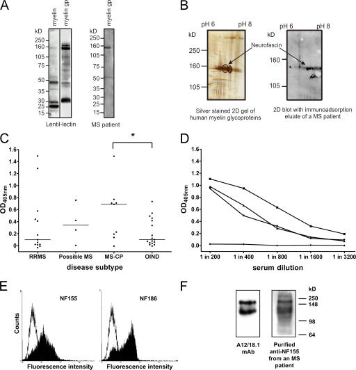
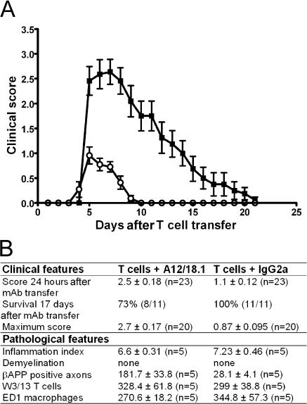
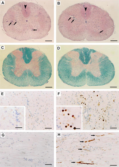
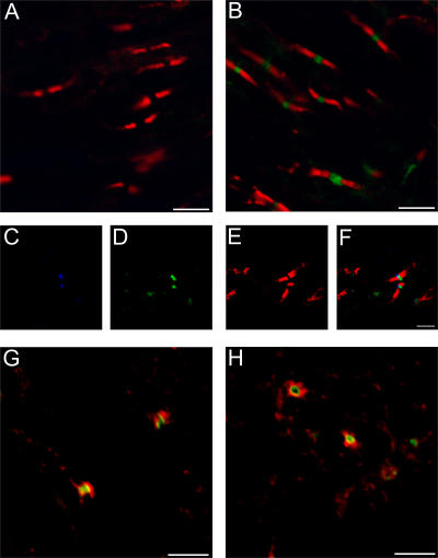
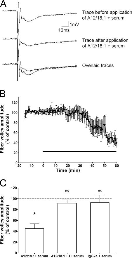
Axonal injury is considered the major cause of disability in patients with multiple sclerosis (MS), but the underlying effector mechanisms are poorly understood. Starting with a proteomics-based approach, we identified neurofascin-specific autoantibodies in patients with MS. These autoantibodies recognize the native form of the extracellular domains of both neurofascin 186 (NF186), a neuronal protein concentrated in myelinated fibers at nodes of Ranvier, and NF155, the oligodendrocyte-specific isoform of neurofascin. Our in vitro studies with hippocampal slice cultures indicate that neurofascin antibodies inhibit axonal conduction in a complement-dependent manner. To evaluate whether circulating antineurofascin antibodies mediate a pathogenic effect in vivo, we cotransferred these antibodies with myelin oligodendrocyte glycoprotein-specific encephalitogenic T cells to mimic the inflammatory pathology of MS and breach the blood-brain barrier. In this animal model, antibodies to neurofascin selectively targeted nodes of Ranvier, resulting in deposition of complement, axonal injury, and disease exacerbation. Collectively, these results identify a novel mechanism of immune-mediated axonal injury that can contribute to axonal pathology in MS.


Figures

Figure 1.
Figure 1.
Antibodies to NF155 and NF186 are present in patients with MS. (A) Human myelin and the myelin glycoprotein (gp) fraction were separated by SDS-PAGE and blotted, and glycoproteins were detected using biotinylated lentil-lectin. The myelin glycoprotein fraction is highly enriched in higher molecular mass glycoproteins (left). Using this myelin glycoprotein fraction to screen patient sera by Western blotting identified antibody responses to a variety of components, including a protein migrating with an apparent molecular mass of ∼155 kD (right). (B) Two two-dimensional gels were run in parallel. One was silver stained (left) and the other was blotted and developed with IgG obtained from an immunoadsorption eluate of an MS patient (right). The encircled spots at 155 kD were excised and identified by MALDI-MS as neurofascin. (C) Serum IgG antibody titers to recombinant NF155ED were measured by ELISA in OIND (n = 17) and MS (n = 26) patients stratified by the disease course. Patients with chronic progressive MS (MS-CP) had the highest median antibody response (horizontal lines) to NF155ED, which was significantly higher than in OIND patients. *, P < 0.05 using the Mann-Whitney U test. (D) The titrations of three sera from high titer MS patients and one negative control donor are shown. (E) Anti-NF155ECD antibodies affinity purified from plasma of an MS patient bind to both isoforms of neurofascin when expressed at the surface of transfected cells (left, NF155; right, NF186), as shown by flow cytometry. The open graph represents the staining of the control cell line, and the shaded graph represents the staining of the neurofascin transfectants. (F) Rat brain homogenate was separated by SDS-PAGE, blotted, and probed with either the neurofascin-specific mAb A12/18.1 or the purified anti-NF155ECD antibodies, indicating recognition of both the 155- and 186-kD isoforms.
Figure 2.
Figure 2.
Passive transfer of antineurofascin antibody exacerbates adoptive transfer EAE. (A) Rats injected with 2 × 10⁶ activated MOGIgD-specific T cells followed by 500 μg IgG2a control antibody on d 4 (open circles) reached a maximum clinical score of 1 ± 0.12 and had completely recovered by day 10 after T cell transfer. Disease activity was exacerbated in rats coinjected with antineurofascin mAb (closed squares). These animals reached a maximum clinical score of 2.7 ± 0.17, and clinical recovery was still incomplete 20 d after T cell transfer. Data are reported as the mean ± SEM. (n = 11). Pooled data from independent experiments are shown. (B) Cotransfer of the NF155/186-specific mAb in rats with adoptively transferred EAE increased disease severity without exacerbating either inflammation or demyelination in the CNS. Neither the inflammatory index (P = 0.25) nor the number of T cells identified with W3/13 (P = 0.84) or the number of macrophages identified with ED1 (P = 0.41) was increased in the animals treated with the NF155/186-specific mAb. In contrast, axonal damage was augmented, as seen by β-APP staining (P = 0.009). Clinical data are pooled from independent experiments. Histological data were obtained from animals killed 48 h after mAb injection. Data are reported as the mean ± SEM. Comparison of groups was statistically evaluated with Mann-Whitney U test.
Figure 3.
Figure 3.
Pathology associated with autoantibody-mediated axonal injury in adoptive transfer EAE. Representative histopathology of the spinal cord 2 d after transfer of either antineurofascin antibody (B, D, F, and H) or an IgG2a control antibody (A, C, E, and G). (A and B) Hematoxylin and eosin staining reveals the presence of similar numbers of inflammatory infiltrates (arrows) in animals coinjected with (A) control mAb and (B) NF155/186-specific mAb. Arrowheads indicate areas in A and B that are shown in immunohistochemical staining for β-APP (E and F) at a higher magnification. (C and D) Luxol fast blue staining for myelin (blue/turquoise) reveals that demyelination is not enhanced by the antineurofascin mAb. In E–H, β-APP is stained brown by immunohistochemistry, and cell nuclei are counterstained blue with hematoxylin. This staining for β-APP, which indicates acute axonal injury, reveals numerous injured axons (brown dots in cross sections, F; brown fibers in longitudinal sections, H) in animals treated with the NF155/186-specific mAb. Note that in comparison with animals treated with the NF155/186-specific mAb (F and H), axonal injury is virtually absent in animals receiving the control IgG2a mAb (E and G). Arrows in H indicate brown β-APP–positive axons. Bars: (A–D) 220 mm; (E–H) 30 mm; (E and F, insets) 13 mm.
Figure 4.
Figure 4.
The panneurofascin-specific mAb A12.18/1 binds selectively to the node of Ranvier in vivo in animals with EAE. Confocal microscopy of representative spinal cord tissue from animals with EAE 30 h after transfer of anti-NF155/NF186 mAb A12.18/1 (B–H) or IgG2a control antibody (A). Cotransfer of anti-NF155/NF186 mAb resulted in deposition of the antibody within discreet regions of the CNS when visualized with an Alexa Fluor 488–conjugated anti–mouse IgG2a antibody (green). Double staining with a rabbit antibody to NF155 (red) identified the paranodal domains of myelinated axons and demonstrated that the injected mAb did not colocalize with NF155 but was deposited between adjacent paranodes (B).There was no deposition of mouse antibody in the CNS of animals injected with the isotype control antibody when visualized with an Alexa Fluor 488–conjugated anti–mouse IgG2a antibody (green), as shown in relation to NF155 staining at the paranode (red, A). Triple staining (C–F) for voltage-gated sodium channels (blue, C), bound anti-NF155/NF186 mAb (green, D), and NF155 (red, E) confirm that the adoptively transferred mAb binds selectively at the node of Ranvier. (F) Merged image of (C–E). Deposition of the in vivo–injected antineurofascin mAb at the node of Ranvier is accompanied by the deposition of complement C9. Sections were stained with an Alexa Fluor 488–conjugated anti–mouse IgG2a antibody to identify bound antineurofascin mAb (green), and a rabbit anti–rat C9 antibody (red, G and H). Bars: 5 μm.
Figure 5.
Figure 5.
Antineurofascin antibody disrupts nerve conduction in vitro. (A) An example of the field potential recorded under control conditions from the CA1 region of a rat hippocampal slice in a single slice using a single stimulus of the Schaffer collateral-commissural fibers every 30 s (top trace) is shown. Perfusion of the antineurofascin mAb A12/18.1 plus normal rat serum causes a decrease in the fiber volley amplitude (middle trace), which can be clearly seen when the traces are overlaid (bottom trace). (B) Time-course data showing the effect of A12/18.1 antibody and serum on the amplitude of the fiber volley (mean ± SEM; n = 5). The horizontal line indicates application of antibody. (C) A decrease in fiber volley amplitude after a 1-h perfusion of the treatment is only seen when A12/18.1 is applied in the presence of fresh rat serum. There is no effect on the fiber volley amplitude if A12/18.1 and heat inactivated (HI) serum are applied or if a control IgG2a antibody and fresh serum are applied (n = 5 for all treatments). The dashed line indicates the control value against which the other values are measured. Significance is determined by a paired t test on the raw (not normalized) data. P = 0.012 for A12/18.1 antibody and fresh serum.

References

    1. Steinman, L. 2001. Multiple sclerosis: a two-stage disease. Nat. Immunol. 2:762–764. - PubMed
    1. Hauser, S.L., and J.R. Oksenberg. 2006. The neurobiology of multiple sclerosis: genes, inflammation, and neurodegeneration. Neuron. 52:61–76. - PubMed
    1. Trapp, B.D., R. Ransohoff, and R. Rudick. 1999. Axonal pathology in multiple sclerosis: relationship to neurologic disability. Curr. Opin. Neurol. 12:295–302. - PubMed
    1. Ferguson, B., M.K. Matyszak, M.M. Esiri, and V.H. Perry. 1997. Axonal damage in acute multiple sclerosis lesions. Brain. 120:393–399. - PubMed
    1. Trapp, B.D., J. Peterson, R.M. Ransohoff, R. Rudick, S. Mork, and L. Bo. 1998. Axonal transection in the lesions of multiple sclerosis. N. Engl. J. Med. 338:278–285. - PubMed

Publication types

MeSH terms