. 2007 Oct;130(4):365-78.
doi: 10.1085/jgp.200709790. Epub 2007 Sep 10.

Triadin binding to the C-terminal luminal loop of the ryanodine receptor is important for skeletal muscle excitation contraction coupling

Affiliations

Triadin binding to the C-terminal luminal loop of the ryanodine receptor is important for skeletal muscle excitation contraction coupling

Sanjeewa A Goonasekera et al. J Gen Physiol. 2007 Oct.

Abstract

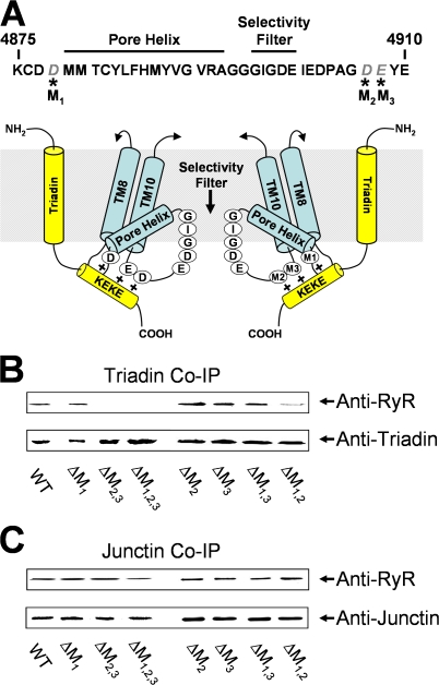
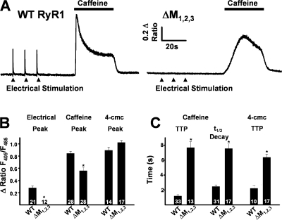
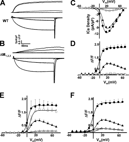
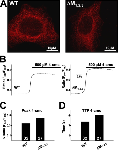
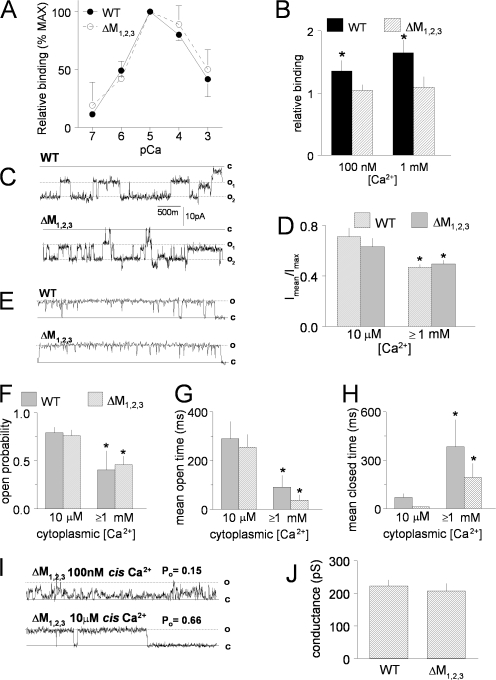
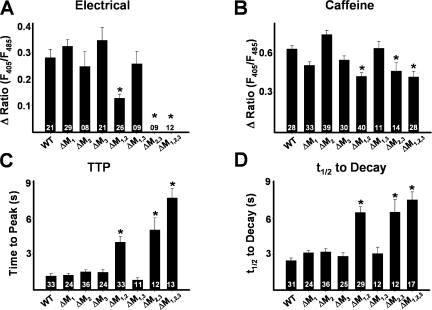
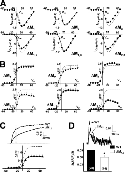
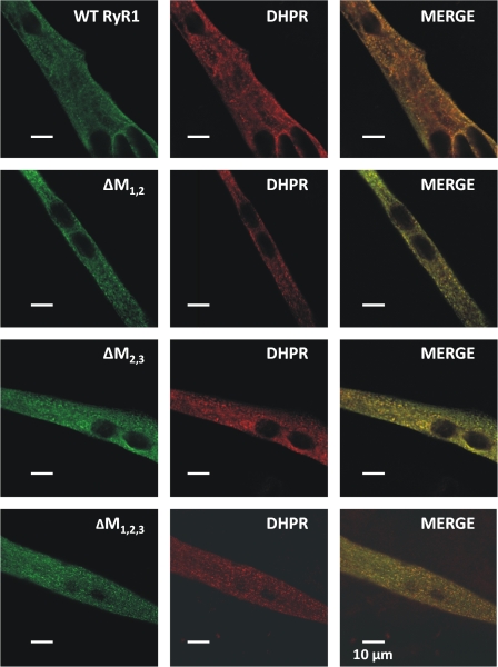
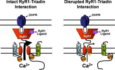
Ca(2+) release from intracellular stores is controlled by complex interactions between multiple proteins. Triadin is a transmembrane glycoprotein of the junctional sarcoplasmic reticulum of striated muscle that interacts with both calsequestrin and the type 1 ryanodine receptor (RyR1) to communicate changes in luminal Ca(2+) to the release machinery. However, the potential impact of the triadin association with RyR1 in skeletal muscle excitation-contraction coupling remains elusive. Here we show that triadin binding to RyR1 is critically important for rapid Ca(2+) release during excitation-contraction coupling. To assess the functional impact of the triadin-RyR1 interaction, we expressed RyR1 mutants in which one or more of three negatively charged residues (D4878, D4907, and E4908) in the terminal RyR1 intraluminal loop were mutated to alanines in RyR1-null (dyspedic) myotubes. Coimmunoprecipitation revealed that triadin, but not junctin, binding to RyR1 was abolished in the triple (D4878A/D4907A/E4908A) mutant and one of the double (D4907A/E4908A) mutants, partially reduced in the D4878A/D4907A double mutant, but not affected by either individual (D4878A, D4907A, E4908A) mutations or the D4878A/E4908A double mutation. Functional studies revealed that the rate of voltage- and ligand-gated SR Ca(2+) release were reduced in proportion to the degree of interruption in triadin binding. Ryanodine binding, single channel recording, and calcium release experiments conducted on WT and triple mutant channels in the absence of triadin demonstrated that the luminal loop mutations do not directly alter RyR1 function. These findings demonstrate that junctin and triadin bind to different sites on RyR1 and that triadin plays an important role in ensuring rapid Ca(2+) release during excitation-contraction coupling in skeletal muscle.


Figures

Figure 1.
Figure 1.
Triadin and junctin binding to RyR1 luminal loop mutants. (A) Proposed interaction between triadin and the terminal RyR1 luminal loop. (Top) Amino acid sequence for a portion of the luminal loop between the final two transmembrane domains of the rabbit RyR1 protein. Negatively charged amino acids mutated in this study (D4878, D4807, and E4809 on the left and M1, M2, and M3 on the right in the schematic) are shown in italics. (Bottom) Representation of the triadin interaction with the terminal luminal loop of RyR1 is modified from Fig. 5 of Lee et al. (2004). (B and C) Western blot analysis of proteins immunoprecipitated with anti-triadin (B) or anti-junctin (C). After incubation of the WT or mutant RyR1 (listed at the bottom of the blot) with either triadin (B) or junctin (C). Immunoprecipitations were performed and the immunoprecipitated protein analyzed by Western Blot using anti-RyR and anti-triadin (B) or anti-junctin (C).
Figure 2.
Figure 2.
Effects of ΔM1,2,3 on ligand-induced Ca2+ release. (A) Representative indo-1 ratio traces obtained from intact dyspedic myotubes expressing either WT RyR1 (left) or ΔM1,2,3 (right) following electrical stimulation (filled triangles) and caffeine application (horizontal bars). (B) Average maximal magnitude of electrically evoked (left), caffeine-induced (middle), and 4-cmc–induced (right) Ca2+ release. (C) Average time to peak (TTP, left) and t 1/2 of decay (middle) for caffeine-induced Ca2+ release and time to peak 4-cmc Ca2+ release (TTP, right). *, P < 0.01.
Figure 3.
Figure 3.
Effects of ΔM1,2,3 on orthograde and retrograde DHPR-RyR1 coupling. (A) Representative L-type Ca2+ currents (bottom traces) and intracellular Ca2+ transients (top traces) resulting from 200-ms depolarizations to −50, −10, +30, and +70 mV in a WT RyR1- expressing myotube. (B) Representative L-type Ca2+ currents (bottom traces) and intracellular Ca2+ transients (top traces) resulting from 200-ms depolarizations to −50, −10, +30, and +70 mV in a ΔM1,2,3-expressing myotube. (C and D) Average voltage dependence of peak L-type Ca2+ current density (C) and intracellular Ca2+ transients (D) in naive dyspedic myotubes (open squares), WT RyR1-expressing (closed circles), and ΔM1,2,3-expressing (closed triangles) myotubes. (E) Inhibition of L-type Ca2+ currents (with 0.5 mM Cd2+/0.2 mM La3+, open symbols) markedly reduced (86 ± 7%, n = 5 at +30 mV) Ca2+ transients in ΔM1,2,3-expressing myotubes (triangles) but only minimally reduced (16 ± 9%, n = 5 at +30 mV) Ca2+ transients in WT RyR1-expressing myotubes (circles). (F) Blockade of Ca2+ release with 100 μM ryanodine (open symbols) markedly reduced depolarization-induced Ca2+ transients in both WT RyR1- (circles) and ΔM1,2,3-expressing (triangles) myotubes.
Figure 4.
Figure 4.
Subcellular localization and Ca2+ release function of WT RyR1 and ΔM1,2,3 channels expressed in HEK293 cells. (A) Both WT RyR1 (left) and ΔM1,2,3 (right) channels exhibit similar reticulated ER expression. (B) Representative fura-2 ratio (F340/F380) traces obtained from WT RyR1- (left) and ΔM1,2,3-expressing (right) HEK293 cells following addition of 500 μM 4-cmc (bar). Average peak (C) and time to peak (TTP) 4-cmc responses (D) obtained from WT RyR1- and ΔM1,2,3-expressing HEK293 cells.
Figure 5.
Figure 5.
Properties of purified WT RyR1 and ΔM1,2,3 channels expressed in HEK293 cells. (A) Ca2+ dependence of [3H]ryanodine binding (% maximum binding) to WT (closed circles) and ΔM1,2,3 (closed squares) channels. (B) Effect of preincubation with 5 μg/ml purified triadin on [3H]ryanodine binding in the presence of either 100 nM or 1 mM Ca2+ (n = 6 for each). *, P < 0.05. [3H]ryanodine binding to purified RyR1 in the presence of triadin (filled bars) is normalized to binding to purified RyR1 in absence of triadin (crosshatched bars). (C) Representative single channel records from artificial lipid bilayers incorporated with two purified WT (top) and two purified ΔM1,2,3 (bottom) channels at −40 mV in the presence of 1 mM trans (luminal) Ca2+ and 10 μM cis (cytoplasmic) Ca2+. The channels opened from the closed level (c) to either single open (o1) and double open (o2) levels. (D) Average data from four WT RyR1 channels and four ΔM1,2,3 channels showing open probability measured as mean current (Imean) normalized to maximum current (Imax) during periods in which only one or two channels were open in the bilayer. Open probability decreased when the cis Ca2+ was increased from 10 μM to either 1 mM or 5 mM (data for 1 and 5 mM cis Ca2+ at +40 and −40 mV were grouped in the average data). *, P < 0.05. (E) Periods of single channel activity in recordings from bilayers containing purified WT (top) and ΔM1,2,3 (bottom) channels at +40 mV with 1 mM trans (luminal) Ca2+ and 10 μM cis (cytoplasmic) Ca2+. (F–H) Average open probability (F), mean open time (G), and mean closed time (H) for 30-s recordings from four WT and four ΔM1,2,3 channels in the presence of 10 μM cis Ca2+ and combined data for 1 and 5 mM cis Ca2+. *, P < 0.05. (I) Data from a ΔM1,2,3 channel recorded first in 100 nM cis Ca2+ (top) and then in 10 μM cis Ca2+ (bottom). The open probability for 30 s of channel activity at each Ca2+ concentration is given above each record. (J) Average single channel conductance from four WT RyR1 channels and four ΔM1,2,3 RyR1 channels.
Figure 6.
Figure 6.
Effects of the terminal luminal loop RyR1 mutants on electrically evoked and caffeine-induced Ca2+ release. (A–D) Bar graphs summarizing the effects of each of WT RyR1 and the different terminal RyR1 luminal loop mutants (ΔM1, ΔM2, ΔM3, ΔM1,2, ΔM1,3, ΔM2,3, and ΔM1,2,3) on electrically evoked Ca2+ release (A), peak caffeine-induced (30 mM) Ca2+ release (B), and the average time to peak (C) and t 1/2 of decay (D) of caffeine-induced Ca2+ transients.
Figure 7.
Figure 7.
Effects of the terminal luminal loop RyR1 mutants on orthograde and retrograde coupling. (A and B) Average voltage dependence of L-type Ca2+ current density (A) and depolarization-induced Ca2+ transients (B) in ΔM1-, ΔM2-, ΔM3-, ΔM1,2-, ΔM1,3-, and ΔM2,3-expressing myotubes. Dashed lines representing the average voltage dependence obtained from WT-expressing myotubes are shown for comparison. (C) Representative depolarization-induced (test potential = +70 mV) Ca2+ transients from WT RyR1- and ΔM1,2-expressing myotubes (top). Voltage dependence of Ca2+ transients measured 30 ms after the start of the test pulse (bottom). (D) Differentials of fluorescence traces (+70 mV) taken during the initial phase of depolarization (top). Bar graph of average peak differential (δ(ΔF/F)/δt) (bottom). The number of experiments is given within each bar. *, P < 0.01.
Figure 8.
Figure 8.
Triadin binding-deficient RyR1 mutants exhibit normal targeting to DHPR-containing junctions. (A) Double immunofluorescence labeling in representative WT- (first row), ΔM1,2- (second row), ΔM2,3- (third row), and ΔM1,2,3-expressing (fourth row) myotubes with antibodies against RyR1 (left) and the DHPR (middle). The “merged images” (right) emphasize common regions of puncta (yellow foci) containing expressed RyR1 proteins and endogenous DHPRs in clusters that represent junctions of the SR with the sarcolemma.
Figure 9.
Figure 9.
Proposed model for triadin regulation of DHPR and ligand activation of RyR1. The quaternary CSQ–triadin–RyR1–junctin interaction tethers CSQ close to the release channel pore and promotes high probability release channel opening (depicted by a large arrow) following either DHPR or ligand activation (left). Disruption of triadin binding to RyR1 results in a similar reduction in SR Ca2+ release (depicted by a small arrow) following either DHPR or ligand activation (right). A junctin–CSQ complex is shown to bind to a separate RyR1 site from that of triadin.

References

    1. Avila, G., J.J. O'Brien, and R.T. Dirksen. 2001. Excitation-contraction uncoupling by a human central core disease mutation in the ryanodine receptor. Proc. Natl. Acad. Sci. USA. 98:4215–4220. - PMC - PubMed
    1. Avila, G., K.M. O'Connell, and R.T. Dirksen. 2003. The pore region of the skeletal muscle ryanodine receptor is a primary locus for excitation-contraction uncoupling in central core disease. J. Gen. Physiol. 121:277–286. - PMC - PubMed
    1. Beard, N.A., M.M. Sakowska, A.F. Dulhunty, and D.R. Laver. 2002. Calsequestrin is an inhibitor of skeletal muscle ryanodine receptor calcium release channels. Biophys. J. 82:310–320. - PMC - PubMed
    1. Beard, N.A., D.R. Laver, and A.F. Dulhunty. 2004. Calsequestrin and the calcium release channel of skeletal and cardiac muscle. Prog. Biophys. Mol. Biol. 85:33–69. - PubMed
    1. Beard, N.A., M.G. Casarotto, L. Wei, M. Varsanyi, D.R. Laver, and A.F. Dulhunty. 2005. Regulation of ryanodine receptors by calsequestrin: effect of high luminal Ca2+ and phosphorylation. Biophys. J. 88:3444–3454. - PMC - PubMed

Publication types

MeSH terms