

Rhabdoid meningioma: clinical features and MR imaging findings in 15 patients
- PMID: 17846191
- PMCID: PMC8134374
- DOI: 10.3174/ajnr.A0601
Rhabdoid meningioma: clinical features and MR imaging findings in 15 patients
Abstract
Background and purpose: Rhabdoid meningioma (RM) is a recently described variant of malignant meningioma, with radiologic features currently not well characterized in the medical literature. The purpose of this study was to describe and characterize clinical features and imaging findings associated with RM.
Materials and methods: CT (n = 8) and MR (n = 15) images of 15 patients (4 men and 11 women; mean age, 52 years; range, 22-75 years) with 16 pathologically proved RMs along with associated clinical records were retrospectively reviewed. All of the patients underwent surgical resection and had additional radiation therapy except for 1 patient. After surgery, the patients had follow-up brain MR imaging to evaluate for tumor recurrence.
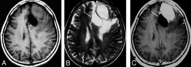
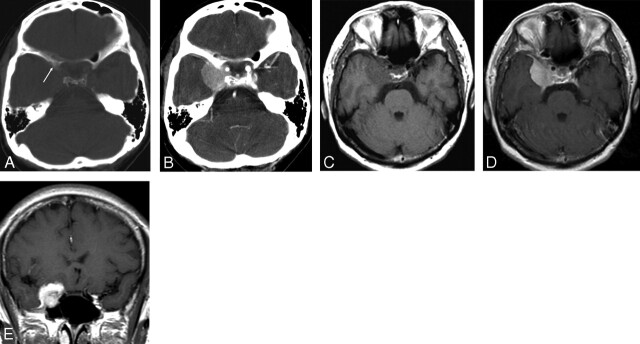
Results: Nine lesions (56%) were located in the cerebral convexity, and 4 lesions (25%) were located in the parasagittal areas. The tumors were isointense (n = 15) to gray matter on T1-weighted images, whereas they were hyperintense (n = 14) on T2-weighted images. On gadolinium-enhanced T1-weighted images, homogeneous enhancement was seen in 10 lesions, and heterogeneous enhancement was seen in 6 lesions that had cysts. Cystic components were noted in 6 lesions (38%). Severe peritumoral edema was seen in 12 lesions (75%). Nine lesions (56%) had hyperostosis, and 5 of them also had bone destruction. Among the 8 cases with initial CT scans, only 1 had amorphous calcifications (13%). There was only 1 recurrence of RM found during the follow-up period after surgical resection.
Conclusion: RMs tend to have prominent peritumoral edema, cystic components, and bone involvement.
Figures
References
-
- Buetow MP, Buetow PC, Smirniotopoulos JG. Typical, atypical, and misleading features in meningioma. Radiographics 1991;11:1087–106 - PubMed
-
- Louis DN, Scheithauer BW, Budka H, et al. Meningiomas. In: Kleihues P, Cavenee WK, eds. World Health Organization Classification of Tumours: Pathology and Genetics of Tumours of the Nervous System. Lyon: IARC Press;2000;176–184
-
- Maier H, Ofner D, Hittmair A, et al. Classic, atypical, and anaplastic meningioma: three histopathological subtypes of clinical relevance. J Neurosurg 1992;77:616–23 - PubMed
-
- Mahmood A, Caccamo DV, Tomecek FJ, et al. Atypical and malignant meningiomas: a clinicopathological review. Neurosurgery 1993;33:955–63 - PubMed
-
- Kolles H, Niedermayer I, Schmitt C, et al. Triple approach for diagnosis and grading of meningiomas: histology, morphometry of Ki-67/Feulgen stainings, and cytogenetics. Acta Neurochir (Wien)1995;137:174–81 - PubMed
MeSH terms
LinkOut - more resources
Full Text Sources
Medical