. 2007 Sep;28(8):1489-93.
doi: 10.3174/ajnr.A0621.

The venous distension sign: a diagnostic sign of intracranial hypotension at MR imaging of the brain

Affiliations

The venous distension sign: a diagnostic sign of intracranial hypotension at MR imaging of the brain

R I Farb et al. AJNR Am J Neuroradiol. 2007 Sep.

Abstract

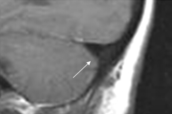
Background and purpose: Patients with intracranial hypotension (IH) demonstrate intracranial venous enlargement with a characteristic change in contour of the transverse sinus seen on routine T1-weighted sagittal imaging. In IH, the inferior margin of the midportion of the dominant transverse sinus acquires a distended convex appearance; we have termed this the venous distension sign (VDS). This is distinct from the normal appearance of this segment, which usually has a slightly concave or straight lower margin. This sign is introduced, and its performance as a test for the presence of this disease is evaluated.

Materials and methods: The transverse sinuses on T1-weighted sagittal imaging of 15 patients with IH and 15 control patients were independently assessed in a blinded fashion by 3 readers for the presence of a VDS. A present or absent VDS was determined for each patient by each reader, and a consensus result for each patient was determined by unanimity or majority rule.

Results: Using the VDS, the readers correctly identified 93% (14 of 15) of the IH patients and similarly 93% (14 of 15) of the control patients. There was a high rate of agreement among the readers for the interpretation of the VDS (multirater kappa = 0.82). The overall sensitivity of the VDS for the diagnosis of intracranial hypotension was 94%. Specificity was also 94%.

Conclusion: The VDS appears to be an accurate test for the presence or absence of IH and may be helpful in the evaluation of these patients.


Figures

Fig 1.
Fig 1.
Normal contour of the transverse sinus. A, T1-weighted image through the approximated middle third of the dominant transverse sinus in a normal control. B, Magnified and cropped image of boxed area in A. C, Same image as B, with schematic representations of the inferior border of the transverse sinus drawn on the image. Both solid and dashed lines represent the normal variability in the contour of this lower border as demonstrated in individual normal controls D through K. The dotted line in C depicts the distended, convex, lower border of the middle third of the dominant transverse sinus (the VDS) as seen in intracranial hypotension (Fig 3).
Fig 2.
Fig 2.
Typical MR findings in IH. A, Midline sagittal T1-weighted image showing the “sagging brain” appearance with distortion of the anterior margin of the pons and medulla (black arrows) and decreased vertical dimension of the suprasellar cistern and sagging tuber cinereum (dashed arrow), as well as the prominent pituitary gland (white arrow). B, Axial T2-weighted image showing effacement of the perimesencephalic cistern (dotted arrows), as well as the elongated anteroposterior dimension of the midbrain. C, Axial T2-weighted gradient echo (susceptibility weighted) image showing small bilateral subdural effusions (short arrows) with a small amount of hemosiderin staining (arrowheads), indicating previous subdural hemorrhage. D, Gadolinium-enhanced axial T1-weighted image demonstrating diffuse pachymeningeal (dural) enhancement (open arrows). E, T1-weighted image through the approximated middle third of the dominant transverse sinus (located by dashed line in B). F, Magnified image of boxed area in E showing the convex inferior margin of the transverse sinus (curved arrow), that is, the VDS indicative of IH.
Fig 3.
Fig 3.
Examples of the VDS in 5 patients with IH pretreatment and posttreatment with EBP. A–E, Magnified and cropped sagittal T1-weighted images through the approximated middle third of the dominant transverse sinus of IHPs 1, 5, 11, 13, and 14 at presentation. A–E′, Corresponding image of each patient at follow-up MR imaging, after EBP and resolution of symptoms. Note the change in the contour of the transverse sinus (particularly the inferior border) between the before and after images of each patient (arrows).
Fig 4.
Fig 4.
False-negative case. Sagittal T1-weighted image through the approximated middle third of the dominant transverse sinus magnified and cropped of IHP 10. This was interpreted as absent VDS by 2 of the 3 readers and was, therefore, registered as a false-negative result. Note the lower margin of the transverse sinus does appear straight (arrow).
Fig 5.
Fig 5.
False-positive case. Sagittal T1-weighted image through the approximated middle third of the dominant transverse sinus magnified and cropped of CP 1. This was interpreted as a VDS by all 3 of the readers and, therefore, registered as a false-positive result. Note that the lower margin of the transverse sinus does appear slightly convex (arrow).

References

    1. Mokri B. Headaches caused by decreased intracranial pressure: diagnosis and management. Curr Opin Neurol 2003;16:319–26 - PubMed
    1. Schievink WI. Spontaneous spinal cerebrospinal fluid leaks and intracranial hypotension. JAMA 2006;295:2286–96 - PubMed
    1. Dillon WP, Fishman RA. Some lessons learned about the diagnosis and treatment of spontaneous intracranial hypotension. AJNR Am J Neuroradiol 1998;19:1001–02 - PubMed
    1. Mokri B. Syndrome of cerebral spinal fluid hypovolemia: clinical and imaging features and outcome. Neurology 2001;56:1607–08 - PubMed
    1. Chung SJ, Kim JS, Lee MC. Syndrome of cerebral spinal fluid hypovolemia: clinical and imaging features and outcome. Neurology 2000;55:1321–27 - PubMed

Publication types