

Distal aneurysms of cerebellar arteries: incidence, clinical presentation, and outcome of endovascular parent vessel occlusion
- PMID: 17846215
- PMCID: PMC8134400
- DOI: 10.3174/ajnr.A0607
Distal aneurysms of cerebellar arteries: incidence, clinical presentation, and outcome of endovascular parent vessel occlusion
Abstract
Background and purpose: The aim of this retrospective study was to report the incidence, clinical presentation, and midterm clinical and imaging results of endovascular parent vessel occlusion of 11 patients with 13 distal cerebellar artery aneurysms.
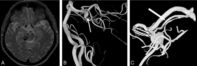
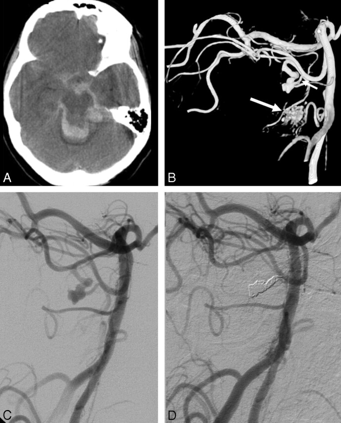
Materials and methods: Between January 1995 and December 2006, 2201 aneurysms were treated in our institution. Thirteen aneurysms in 11 patients were located on distal cerebellar arteries (incidence, 0.6%), 8 of them arising from vessels feeding small arteriovenous malformations. There were 6 men and 5 women, ranging from 44 to 70 years of age. One patient with a superior cerebellar artery aneurysm presented with isolated trochlear nerve palsy. Ten patients presented with subarachnoid and intraventricular hemorrhage, and most patients were in poor clinical condition on admission. Aneurysm location was the superior cerebellar artery in 3, the anterior inferior cerebellar artery in 5, and the posterior inferior cerebellar artery in 5. Two patients had 2 aneurysms each.
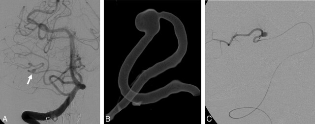
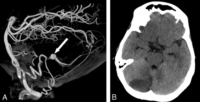
Results: Eleven aneurysms were treated by simultaneous coil occlusion of the aneurysm and parent artery or occlusion of the parent artery just proximal to the aneurysm. Clinical follow-up was at a mean of 16.5 months (range, 2-40 months). Infarction in the territory of the occluded vessel was apparent on follow-up imaging in 5 of 11 patients, all without functional impairment.
Conclusion: Distal cerebellar artery aneurysms are rare. Most patients present with poor-grade hemorrhage. Endovascular parent vessel occlusion is effective in excluding the aneurysm from the circulation. In most patients, adequate collateral circulation prevents infarction in the territory of the occluded vessel. In this series, when infarction did occur, the clinical consequences were limited.
Figures
References
-
- Locksley HB. Natural history of subarachnoid hemorrhage, intracranial aneurysms and arteriovenous malformations: based on 6368 cases in the cooperative study. J Neurosurg 1966;25:215–39 - PubMed
Publication types
MeSH terms
LinkOut - more resources
Full Text Sources
Medical