

A new covered stent designed for intracranial vasculature: application in the management of pseudoaneurysms of the cranial internal carotid artery
- PMID: 17846216
- PMCID: PMC8134415
- DOI: 10.3174/ajnr.A0668
A new covered stent designed for intracranial vasculature: application in the management of pseudoaneurysms of the cranial internal carotid artery
Abstract
Background and purpose: The management of intracranial pseudoaneurysms is controversial. The purpose of this study was to provide a preliminary evaluation of the clinical efficacy of a Willis covered stent specially designed for the intracranial vasculature in the management of a pseudoaneurysm of the cranial internal carotid artery (CICA).
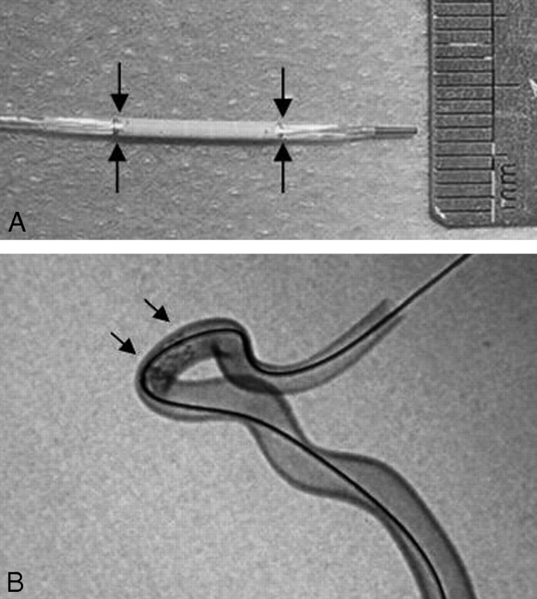
Materials and methods: Eight patients with pseudoaneurysms of the CICA were treated with use of the Willis covered stent. The flexibility of the entire stent system was gauged from the resistance met when reaching the target lesion and was categorized as no resistance, no apparent resistance, or resistance that could be overcome. The apposition of the Willis stent after deployment was scored as excellent with no endoleak, good with a small endoleak, or bad with an apparent endoleak. Follow-up angiography was performed 3 to 12 months after placement of the stent, and angiographic assessments were categorized as endoleak, stenosis of the covered segment of vessel, or occlusion of parent arteries. Follow-up clinical evaluations were also performed, and outcomes were graded as full recovery, improvement, unchanged, and aggravation.
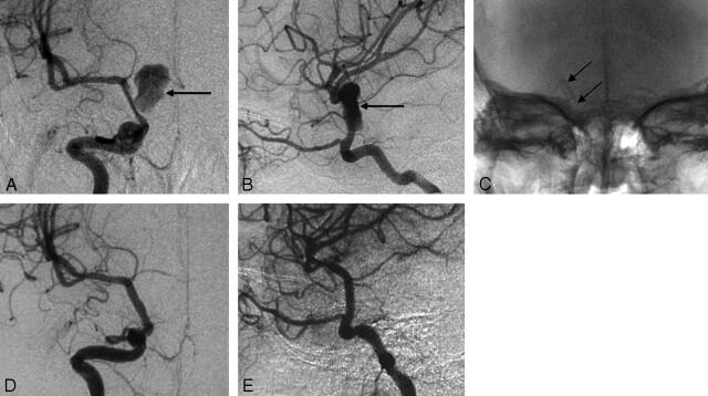
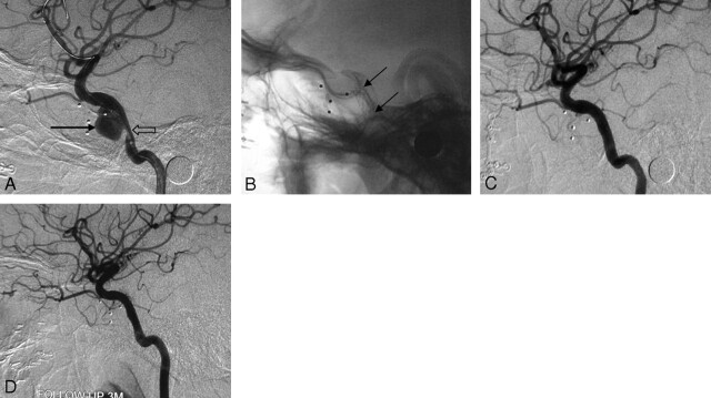
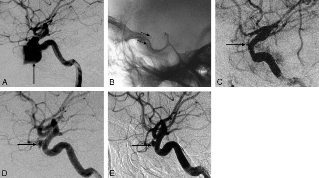
Results: Endovascular treatment was technically successful in all aneurysms without procedural-related complications, and all of the stents were easily navigated to the targeted lesions in the CICA. Complete resolution of the pseudoaneurysm was observed in 6 patients immediately after the procedure, and a minimal endoleak into the aneurysm persisted in 2 patients. No morbidity or mortality and no technical adverse event occurred. A follow-up angiogram confirmed complete reconstruction of the internal carotid artery, with no recurrent aneurysmal filling and no occurrence of stenosis in the area of the stent. By the final follow-up visit, 4 patients had fully recovered, 3 had improved, and 1 patient's condition was unchanged.
Conclusion: On the basis of our preliminary experience, the Willis covered stent specially designed for the intracranial vasculature can manage a CICA pseudoaneurysm safely and effectively, but longer follow-up and expanded clinical trials are needed.
Figures
References
-
- Biondi A. Intracranial aneurysms associated with other lesions, disorders or anatomic variations. Neuroimaging Clin N Am 2006;16:467–82, viii - PubMed
-
- Alexander MJ, Smith TP, Tucci DL. Treatment of an iatrogenic petrous carotid artery pseudoaneurysm with a Symbiot covered stent: technical case report. Neurosurgery 2002;50:658–62
-
- Biffl WL, Moore EE, Offner PJ, et al. Blunt carotid arterial injuries: implications of a new grading scale. J Trauma 1999;47:845–53 - PubMed
-
- Kadyrov NA, Friedman JA, Nichols DA, et al. Endovascular treatment of an internal carotid artery pseudoaneurysm following transsphenoidal surgery. Case report. J Neurosurg 2002;96:624–27 - PubMed
Publication types
MeSH terms
Substances
LinkOut - more resources
Full Text Sources
Research Materials
Miscellaneous