

Case Reports
doi: 10.3174/ajnr.A0606.
Dural arteriovenous fistula involving the posterior condylar canal
Affiliations
- PMID: 17846219
- PMCID: PMC8134384
- DOI: 10.3174/ajnr.A0606
Item in Clipboard
Case Reports
Dural arteriovenous fistula involving the posterior condylar canal
AJNR Am J Neuroradiol.
2007 Sep.
Abstract
Although dural arteriovenous fistulas (DAVFs) occur in any structure that is covered by the dura mater, DAVFs at the posterior condylar canal have not been reported. We present a DAVF that involves the posterior condylar canal and drains into the posterior condylar vein and the occipital sinus, which was treated by selective transvenous embolization. Knowledge of venous anatomy of the craniocervical junction and careful assessment of the location of the arteriovenous fistula can contribute to successful treatment.
Figures
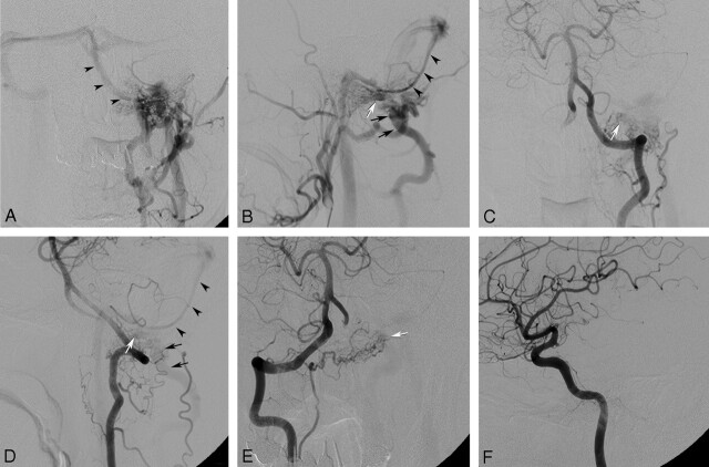
Frontal (A) and lateral (B) views of an angiogram of the left external carotid artery show the AVF (white arrow) being fed by the left ascending pharyngeal artery and the left occipital artery, draining through the posterior condylar vein (black arrows) into the posterior cervical vein and sigmoid sinus and into the occipital sinus (arrowheads). Frontal (C) and lateral (D) views of an angiogram of the left vertebral artery show the AVF (white arrow) being fed by the left anterior meningeal artery. Black arrows indicate the posterior condylar vein, and arrowheads indicate the occipital sinus. Angiogram of the right vertebral artery (E) demonstrates the shunted venous pouch (white arrow) being fed by the right anterior meningeal artery. Angiogram of the left internal carotid artery (F) shows the AVF being fed by the dorsal clival artery.
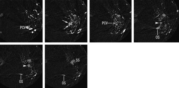
Axial reconstructed images of rotational angiogram of the left external carotid artery show the fistulous pouch (white arrows) draining through the posterior condylar vein (PCV) into the posterior cervical vein inferiorly and the occipital sinus (OS) posterosuperiorly. The occipital sinus and the posterior condylar vein form a common trunk (arrowheads) that joins into the sigmoid sinus (SS).
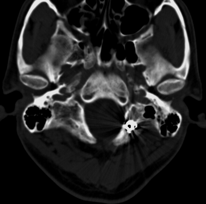
CT after embolization shows coils at the left posterior condylar canal.
References
-
- Takahashi S, Sakuma I, Omachi K, et al. Craniocervical junction venous anatomy around the suboccipital cavernous sinus: evaluation by MR imaging. Eur Radiol 2005;15:1694–700 - PubMed
-
- Kiyosue H, Tanoue S, Okahara M, et al. Ocular symptoms associated with a dural arteriovenous fistula involving the hypoglossal canal: selective transvenous coil embolization. Case report. J Neurosurg 2001;94:630–32 - PubMed
Publication types
MeSH terms
LinkOut - more resources
Full Text Sources