Review
. 2007 Oct;8(10):776-90.
doi: 10.1038/nrg2172. Epub 2007 Sep 11.

Ribozymes, riboswitches and beyond: regulation of gene expression without proteins

Affiliations
Review

Ribozymes, riboswitches and beyond: regulation of gene expression without proteins

Alexander Serganov et al. Nat Rev Genet. 2007 Oct.

Abstract

Although various functions of RNA are carried out in conjunction with proteins, some catalytic RNAs, or ribozymes, which contribute to a range of cellular processes, require little or no assistance from proteins. Furthermore, the discovery of metabolite-sensing riboswitches and other types of RNA sensors has revealed RNA-based mechanisms that cells use to regulate gene expression in response to internal and external changes. Structural studies have shown how these RNAs can carry out a range of functions. In addition, the contribution of ribozymes and riboswitches to gene expression is being revealed as far more widespread than was previously appreciated. These findings have implications for understanding how cellular functions might have evolved from RNA-based origins.


Conflict of interest statement

Competing interests statement

The authors declare no competing financial interests.

Figures
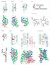
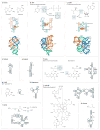
Figure 1
Figure 1. Domain organization and secondary and three-dimensional structures of ribozymes
Secondary structures are depicted in thick lines and are connected by thin black lines with arrows. Watson–Crick and non-canonical base pairs are shown as solid lines and circles, respectively. Bulged-out nucleotides are represented as triangles. Ribozymes with known three-dimensional structures are coloured according to secondary structure elements and domains. Three-dimensional structures, if available, are shown in a ribbon-and-stick representation below the secondary structures. The two nucleotides that lie adjacent to the scissile phosphate are indicated by red boxes. Nucleotides that are implicated in catalysis (panels af and i) or that are essential for molecular recognition (panel j) are indicated by yellow boxes. Black dashed squares and lines highlight important tertiary interactions. Coloured dashed lines indicate elements that are missing in the structure or that are substituted by non-natural sequences. a | Hammerhead ribozyme. b | Hairpin ribozyme. c | Hepatitis δ virus ribozyme. d | Varkud satellite (VS) ribozyme. e | CPEB3 ribozyme. f | Human CoTC ribozyme. g | Bacillus antracis glmS ribozyme; glucosamine-6-phosphate (GlcN6P) is represented as a red oval, with its interactions with RNA indicated by dashed lines. h | Bacillus subtilis RNase P, B-type. i | Domain organization of the group II intron, with IBS and EBS designating intron and exon binding sequences, respectively,. The yellow-coloured A designates a conserved unpaired adenosine that participates in splicing. j | Asoarcus spp. BH72 group I intron in the state that precedes the second step of splicing. The internal guide sequence (IGS) aligns the 5′ and 3′ exons (ex), which are shown in grey. ωG and αG designate the 3′-terminal guanosine nucleotide of the intron and the external guanosine that is linked to the intron after the first step of splicing, respectively. Secondary structures in panels a, c, d, e, f, g, h and j are modified with permission from REF. © (2006) Cell Press, REF. © (2007) Cell Press, REF. © (1995) National Academy of Sciences (USA), REF. © (2006) American Association for the Advancement of Science, Nature REF. © (2004) Macmillan Publishers Ltd, REF. © (2006) American Association for the Advancement of Science and REF. © (2007) Current Biology Ltd, REF. © (2006) Elsevier Sciences and REF. © (2005) Elsevier Sciences, respectively.
Figure 2
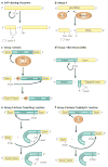
Figure 2. Reactions catalysed by ribozymes
a | The typical reaction of self-cleaving ribozymes, initiated by 2′-hydroxyl (OH) attack, and yielding 2′,3′-cyclic phosphate (P) and 5′-OH termini. b | The catalytic cleavage of pre-tRNA by RNase P (REF. 2). A water molecule serves as a nucleophile, and the reaction yields 2′,3′-diol and 5′-P termini. c | Self-splicing by group I introns. The reaction is initiated by nucleophilic attack by the 3′-OH of external guanosine (αG) at the 5′ splice site. This results in covalent linkage of αG to the 5′ end of the intron and release of the 3′-OH of the 5′ exon. In the second step, the 3′-OH attacks the 3′ splice site located immediately after the conserved guanosine (ωG), resulting in excision of the intron with αG at the 5′ end and release of the ligated exons. d | The ‘capping’ reaction of the Didium iridis GIR1 ribozyme is similar to the first step of the ‘branching’ reaction of group II introns. The reaction joins nucleotides by a 2′,5′-phosphodiester linkage, thereby forming a 3-nucleotide ‘lariat’ that might be a protective 5′ cap of the mRNA. e | The self-splicing of group II introns by a branching reaction. In the first step, the 5′ splice site is attacked by the 2′-OH of a conserved unpaired adenosine located in domain VI, resulting in formation of a 2′,5′-phosphodiester linkage. In the next step, the free 3′-OH group of the 5′ exon attacks the 3′ splice site, liberating the circular intron lariat and ligated exons. f | Alternative ‘hydrolytic’ self-splicing of group II introns. The reaction involves a water molecule as a nucleophile and produces a linear intron.
Figure 3
Figure 3. Gene regulation by RNA switches
RNA regions that are involved in gene expression switching are shown in the same colour. a | Translation activation of virulence genes in the pathogen Listeria monocytogenes. An increase in temperature melts the secondary structure around the ribosome binding site (RBS) and start codon, allowing ribosome binding and translation initiation. b | Upregulation of Escherichia coli σs-factor gene by the DsrA antisense short RNA (sRNA). DsrA RNA pairs with the translational operator of the rpoS gene using two sequences (coloured blue and light blue) located within helices 1 and 2 (REFs 33,34,121). This base pairing exposes translation initiation signals for ribosome binding and increases mRNA stability. c | Downregulation of transcription regulator HNS by DsrA sRNA. DsrA RNA, transcribed in response to low temperature, pairs with 5′ and 3′ regions of hns mRNA and causes faster turnover of the mRNA, possibly by RNase E degradation. d | Transcription termination of the Bacillus subtilis glycyl-tRNA synthetase gene by aminoacylated tRNAGly (REF. 124). Non-aminoacylated tRNAGly interacts with the T-box region of mRNA using an anticodon and an acceptor helix, and promotes the formation of the anti-terminator stem–loop structure. The aminoacylated tRNA cannot contact mRNA using the acceptor stem, thus allowing the formation of the transcription terminator. e | Transcription activation of the purine efflux pump by the adenine riboswitch. In the absence of adenine, transcription of B. subtilis ydhL mRNA is aborted as a result of formation of a transcription terminator. Adenine binding stabilizes the metabolite-sensing domain and prevents the formation of the terminator. f | Thiamine pyrophosphate (TPP)-riboswitch-mediated alternative splicing of mRNA in Neurospora crassa. In the absence of TPP, the mRNA adopts a structure that occludes the 5′ splice site by base pairing with the P4–P5 region of the riboswitch. Pre-mRNA splicing from 5′ splice site 1 leads to production of a short mRNA and expression of the NMT1 gene. TPP binding causes a structural change in the RNA, opening the 5′ splice site 2 and occluding the branch site. Therefore, splicing is inhibited (not shown) and, were it to proceed, would result in the formation of a long mRNA. The alternatively spliced and non-spliced mRNAs both carry a short ORF (μORF) that begins from initiation codons 1–2 and competes with translation of the main ORF, thereby repressing NMT1 expression. Key splicing determinants are activated (indicated by green arrows) and inhibited (indicated by red lines) during different occupancy states of the TPP-sensing domain. Panels a, b and c, d and f are modified with permission from REF. © (2002) Cell Press, REF. © (2000) National Academy of Sciences (USA), REF. © (2005) Academic Press and Nature REF. © (2007) Macmillan Publishers Ltd.
Figure 4
Figure 4. Secondary and tertiary structures of riboswitches
Secondary structures are depicted by thick lines and are connected by black lines with arrows. Watson–Crick and non-canonical base pairs are shown as solid lines and circles, respectively. Natural riboswitch ligands and riboswitch-binding antibiotics are shown next to the secondary structures. Grey shadings indicate areas in which the ligands undergo changes. a | The Bacillus subtilis xpt gene guanine riboswitch bound to guanine (represented as a red G). The discriminatory nucleotide C74 is coloured yellow. b | The TPP riboswitch from the Escherichia coli thiM gene bound with TPP (in red). A pair of hydrated Mg2+ cations is shown in magenta. G40 interacting with the pyrimidine moiety of TPP is shown in yellow. The antibiotic pyrithiamine pyrophosphate (PTPP) differs from TPP by the central ring. c | The class I SAM Thermoanaerobacter tengcongensis riboswitch in complex with SAM (in red). U57 interacting with the purine moiety of SAM is shown in yellow. The grey area highlights a methyl group that is missing in S-adenosylhomocysteine (SAH). d | A class II SAM riboswitch from the Agrobacterium tumefaciens metA gene. e | An SMK (class III SAM) riboswitch from the Streptococcus gordonii metK gene. Helix P3 is formed by Shine–Dalgarno and anti-Shine–Dalgarno sequences. f | The FMN riboswitch from the B. subtilis ribD gene. g | The preQ1 riboswitch from the B. subtilis queC gene. h | The magnesium riboswitch from the Salmonella enterica mgtA gene. i | The lysine riboswitch from the B. subtilis lysC gene. j | The AdoCbl riboswitch from the E. coli btuB gene. k | The glycine type II riboswitch from Vibrio cholerae gcvT gene. Secondary structures in panels c, d, e, f, g, h, i, j and k modified with permission from Nature REF. © (2006) Macmillan Publishers Ltd, REF. © (2005) BioMed Central Ltd, Nature Structural & Molecular Biology REF. © (2006) Macmillan Publishers Ltd, REF. © (2002) National Academy of Sciences (USA), Nature Structural & Molecular Biology REF. © (2007) Macmillan Publishers Ltd, REF. © (2006) Cell Press, Nature Biotechnology REF. © (2006) Macmillan Publishers Ltd, REF. © (2002) Current Biology Ltd and REF. © (2004) American Association for the Advancement of Science, respectively.

Similar articles

Cited by

References

    1. Cech TR, Zaug AJ, Grabowski PJ. In vitro splicing of the ribosomal RNA precursor of Tetrahymena: involvement of a guanosine nucleotide in the excision of the intervening sequence. Cell. 1981;27:487–496. - PubMed
    1. Guerrier-Takada C, Gardiner K, Marsh T, Pace N, Altman S. The RNA moiety of ribonuclease P is the catalytic subunit of the enzyme. Cell. 1983;35:849–857. - PubMed
    1. Gilbert W. Origin of life: the RNA World. Nature. 1986;319:618.
    1. Mironov AS, et al. Sensing small molecules by nascent RNA: a mechanism to control transcription in bacteria. Cell. 2002;111:747–756. References 4–6 describe the discovery of metabolite-binding riboswitches. - PubMed
    1. Nahvi A, et al. Genetic control by a metabolite binding mRNA. Chem Biol. 2002;9:1043–1049. - PubMed