

Area extraction of the liver and hepatocellular carcinoma in CT scans
- PMID: 17846836
- PMCID: PMC3043880
- DOI: 10.1007/s10278-007-9053-4
Area extraction of the liver and hepatocellular carcinoma in CT scans
Abstract
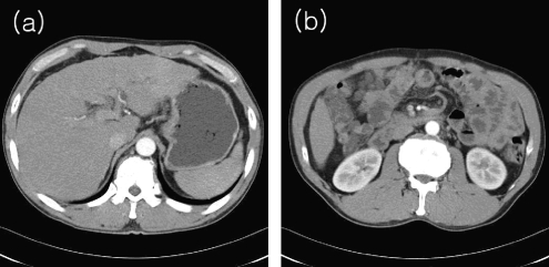
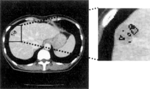
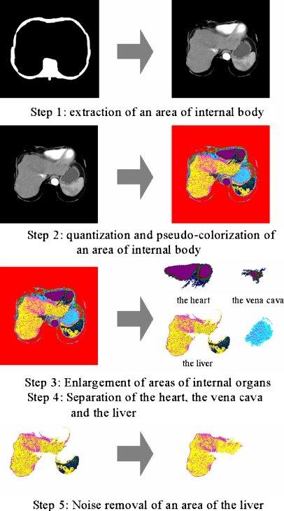
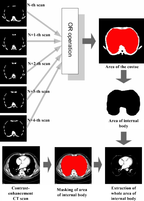
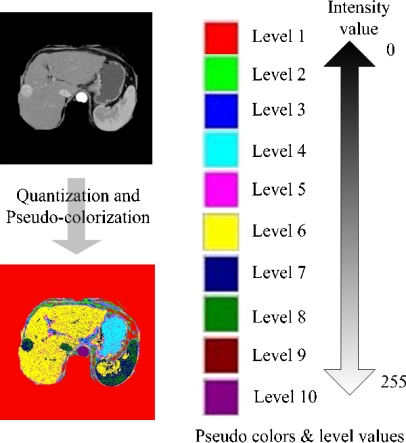
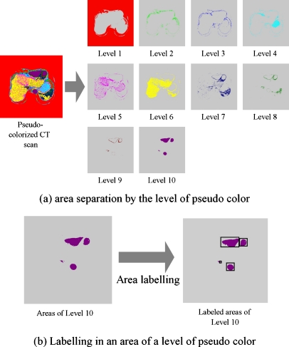
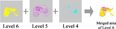
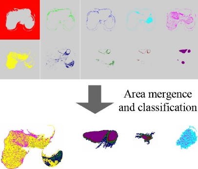
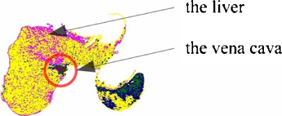
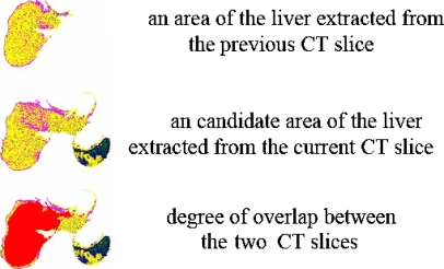
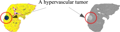
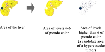
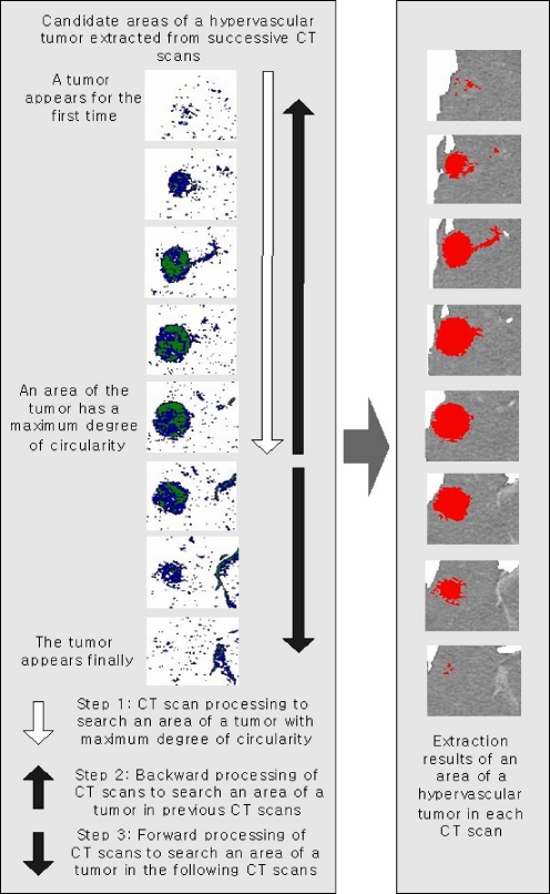
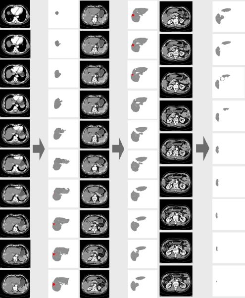
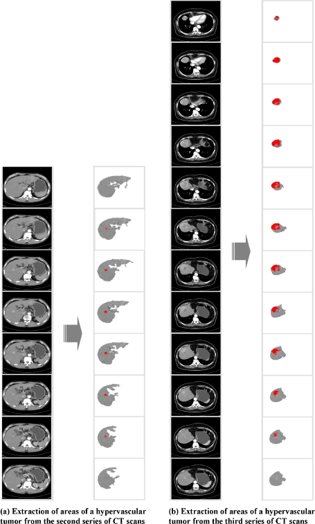
In Korea, hepatocellular carcinoma is the third frequent cause of cancer death, occupying 17.2% among the whole deaths from cancer, and the rate of death from hepatocellular carcinoma comes to about 21 out of 100,000. This paper proposes an automatic method for the extraction of areas being suspicious as hepatocellular carcinoma from computed tomography (CT) scans and evaluates the availability as an auxiliary tool for the diagnosis of hepatocellular carcinoma. For detecting tumors in the internal of the liver from a CT scan, first, an area of the liver is extracted from about 45-50 CT slices obtained by scanning in 2.5-mm intervals starting from the lower part of the chest. In the extraction of an area of the liver, after the unconcerned areas outside of the bony thorax are removed, areas of the internal organs are segmented by using information on the intensity distribution of each organ, and an area of the liver is extracted among the segmented areas by using information on the position and morphology of the liver. Because hepatocellular carcinoma is a hypervascular tumor, the area corresponding to hepatocellular carcinoma appears more brightly than the surroundings in a CT scan, and also takes a spherical shape if the tumor shows expansile growth pattern. By using these features, areas being brighter than the surroundings and globe-shaped are segmented as candidate areas for hepatocellular carcinoma in the area of the liver, and then, areas appearing at the same position in successive CT slices among the candidates are discriminated as hepatocellular carcinoma. For the performance evaluation of the proposed method, experimental results obtained by applying the proposed method to CT scans were compared with the diagnoses by radiologists. The evaluation results showed that all areas of the liver and hypervascular tumors were extracted exactly and the proposed method has a high availability as an auxiliary diagnosis tool for the discrimination of liver tumors.
Figures
Similar articles
-
Small hypervascular hepatocellular carcinoma revealed by double arterial phase CT performed with single breath-hold scanning and automatic bolus tracking.AJR Am J Roentgenol. 2002 Apr;178(4):899-904. doi: 10.2214/ajr.178.4.1780899. AJR Am J Roentgenol. 2002. PMID: 11906869
-
CT diagnosis of early hepatocellular carcinoma: sensitivity, findings, and CT-pathologic correlation.AJR Am J Roentgenol. 1995 Apr;164(4):885-90. doi: 10.2214/ajr.164.4.7726041. AJR Am J Roentgenol. 1995. PMID: 7726041
-
Visualization of hypervascular liver lesions During TACE: comparison of angiographic C-arm CT and MDCT.AJR Am J Roentgenol. 2008 Apr;190(4):W263-9. doi: 10.2214/AJR.07.2695. AJR Am J Roentgenol. 2008. PMID: 18356419
-
Multidetector row CT and MR imaging in diagnosing hepatocellular carcinoma.Intervirology. 2004;47(3-5):209-26. doi: 10.1159/000078474. Intervirology. 2004. PMID: 15383731 Review.
-
Hepatocellular carcinoma in advanced liver cirrhosis: CT detection in transplant patients.Abdom Imaging. 2004 Mar-Apr;29(2):203-7. doi: 10.1007/s00261-003-0114-y. Abdom Imaging. 2004. PMID: 15290946 Review.
Cited by
-
An automated liver tumour segmentation from abdominal CT scans for hepatic surgical planning.Int J Comput Assist Radiol Surg. 2018 Aug;13(8):1169-1176. doi: 10.1007/s11548-018-1801-z. Epub 2018 Jun 2. Int J Comput Assist Radiol Surg. 2018. PMID: 29860549
References
-
- Nakagawa J, Shimizu A, Kobatake H. Development of an automated extraction method for liver tumors in three-dimensional multiphase multislice images. Syst Comput Jpn. 2005;36(9):43–54. doi: 10.1002/scj.20179. - DOI
-
- Henkei RD: Segmentation in scale space. Proceedings of Computer Analysis of Images and Pattern, CAIP, Prague, 1995
Publication types
MeSH terms
Substances
LinkOut - more resources
Full Text Sources
Medical
