. 2007 Oct;81(4):792-8.
doi: 10.1086/521275. Epub 2007 Aug 31.

A defect in the ionotropic glutamate receptor 6 gene (GRIK2) is associated with autosomal recessive mental retardation

Affiliations

A defect in the ionotropic glutamate receptor 6 gene (GRIK2) is associated with autosomal recessive mental retardation

Mohammad Mahdi Motazacker et al. Am J Hum Genet. 2007 Oct.

Abstract

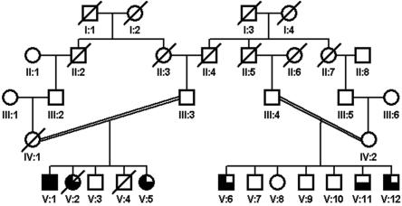
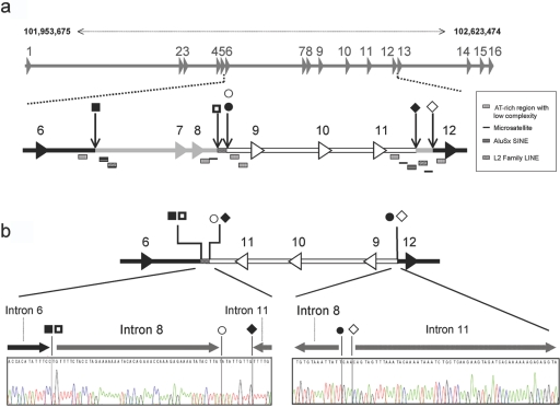
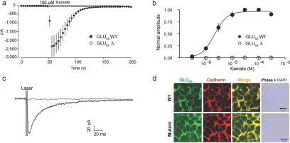
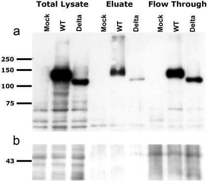
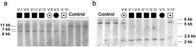
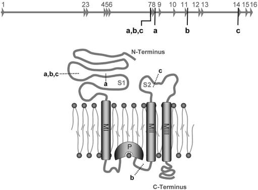
Nonsyndromic mental retardation is one of the most important unresolved problems in genetic health care. Autosomal forms are far more common than X-linked forms, but, in contrast to the latter, they are still largely unexplored. Here, we report a complex mutation in the ionotropic glutamate receptor 6 gene (GRIK2, also called "GLUR6") that cosegregates with moderate-to-severe nonsyndromic autosomal recessive mental retardation in a large, consanguineous Iranian family. The predicted gene product lacks the first ligand-binding domain, the adjacent transmembrane domain, and the putative pore loop, suggesting a complete loss of function of the GLU(K6) protein, which is supported by electrophysiological data. This finding provides the first proof that GLU(K6) is indispensable for higher brain functions in humans, and future studies of this and other ionotropic kainate receptors will shed more light on the pathophysiology of mental retardation.


Figures

Figure  1.
Figure 1.
Family pedigree. Blackened symbols represent patients with severe MR, three-quarters-blackened symbols represent patients with moderate MR, and half-blackened symbols represent patients with mild MR.
Figure  2.
Figure 2.
Genomic mutation in GRIK2. a, Schematic representation of the GRIK2 wild-type allele. Physical coordinates (top) are according to NCBI build 36.1. Exons are depicted by numbered arrowheads. Black shading indicates unaffected sequence, gray shading indicates deleted regions, and the inversion is white outlined in black. A checkered pattern marks the uninverted sequence fragment 5′ of the deletion of exons 7 and 8. Squares, circles, and diamonds mark the positions of the deletion and inversion borders. Rectangular symbols below the schematic of GRIK2 mark the locations of regions of low sequence complexity or repetitive elements. b, Schematic representation of the GRIK2 mutant allele and the sequences of the 5′ and 3′ genomic junction fragments. LINE = long interspersed nuclear element; SINE = short interspersed nuclear element.
Figure  3.
Figure 3.
Loss of function of GLUK6Δ. a, Mean currents evoked by 100 μM kainate (for GLUK6 wild type [WT], n=8; for GLUK6Δ, n=9). b, Dose-response curve for kainate, normalized to response in 100 μM kainate. Error bars represent SEM (for GLUK6WT, n=6–8 per reading point; for GLUK6Δ, n=3–9 per reading point). c, Cells expressing GLUK6WT show photolytically evoked glutamate responses without prior incubation in concanavalin A (black line), whereas GLUK6Δ-expressing cells show no currents after fast glutamate application (gray line). Each trace represents the mean of five successive sweeps. d, Immunocytochemistry showing colocalization of both GLUK6 WT and GLUK6Δ with the endogenous plasma membrane protein cadherin. DAPI = 4′,6-diamidino-2-phenylindole.
Figure  4.
Figure 4.
GLUK6Δ present on the plasma membrane of transfected HEK293 cells. a, Membrane proteins biotinylated on HEK293 cells overexpressing GLUK6 wild type (WT) or GLUK6Δ (Delta) at 48 h after transfection (Pinpoint [Pierce]). Nontransfected HEK293 cells served as a control (Mock). After cell lysis (Total Lysate samples), biotinylated membrane proteins were bound to a NeutrAvidin affinity column. Nonbiotinylated protein was separated, because it does not bind to the NeutrAvidin affinity column (Flow Through samples). Membrane proteins were eluted with sample buffer (Eluate samples). All fractions were separated on a 10% SDS gel, were blotted, and were probed for GLUK6 (with 05-921 [Upstate Biotechnology] at 1:800). GLUK6 WT and GLUK6Δ are present in the membrane protein fraction (Eluate samples). b, Glyceraldehyde 3-phosphate dehydrogenase (GAPDH) loading control (ab9482 [Abcam]).
Figure  5.
Figure 5.
Southern blots for patients and heterozygous carriers of the mutation, characterizing the 5′ border (a) (by use of BglII) and the 3′ border (b) (by use of EcoRV) of the mutation.
Figure  6.
Figure 6.
Impact of GRIK2 mutation on GLUK6 protein structure. Shown is a schematic representation of the GRIK2 wild-type allele and a cartoon of a GLUK6 monomer. MI–MIII = transmembrane domains; P = pore loop. Letters indicate the start and end of different alterations in the genomic sequence and their corresponding impact on the resulting protein. a = deletion of exons 7 and 8; b = complete genomic rearrangement, as observed in patients with MR; c = region lacking in the putative transcript with a junction between exons 6 and 14.
Figure  7.
Figure 7.
Array CGH results for patients with MR. Submegabase-resolution array CGH was performed as described elsewhere. After normalization by subgrid LOWESS, Cy3:Cy5 intensity ratios of each clone were plotted in a size-dependent manner along the chromosome ideograms, by use of CGHPRO. Red and green bars indicate the log2 ratio thresholds of −0.3 (loss) and 0.3 (gain), respectively. A homozygous deletion of 6q16.3 was the only aberration that cosegregated with the disease; it was present in all affected probands and was absent in all unaffected probands tested. Array CGH data shown here have been deposited in NCBI Gene Expression Omnibus (GEO) and are accessible through GEO series accession number GSE 7886. a, Close-up of findings for patient 6q16.3, demonstrating a deletion of two BAC clones (RP11-226C08 and RP11-543M18 [arrows]) encompassing GRIK2. (Because of lower hybridization quality for V:6, only one BAC represents the DNA copy-number loss.) b, Overall view of chromosome 6 in the corresponding patients. A red rectangle highlights the region depicted in panel a.

References

Web Resource

    1. Online Mendelian Inheritance in Man (OMIM), http://www.ncbi.nlm.nih.gov/Omim/ (for neurotrypsin, cereblon, CC2D1A, MRT6, GRIK2, and fragile X syndrome)

References

    1. Molinari F, Rio M, Meskenaite V, Encha-Razavi F, Auge J, Bacq D, Briault S, Vekemans M, Munnich A, Attié-Bitach T, et al (2002) Truncating neurotrypsin mutation in autosomal recessive nonsyndromic mental retardation. Science 298:1779–178110.1126/science.1076521 - DOI - PubMed
    1. Higgins JJ, Pucilowska J, Lombardi RQ, Rooney JP (2004) A mutation in a novel ATP-dependent Lon protease gene in a kindred with mild mental retardation. Neurology 63:1927–1931 - PMC - PubMed
    1. Basel-Vanagaite L, Attia R, Yahav M, Ferland RJ, Anteki L, Walsh CA, Olender T, Straussberg R, Magal N, Taub E, et al (2006) The CC2D1A, a member of a new gene family with C2 domains, is involved in autosomal recessive non-syndromic mental retardation. J Med Genet 43:203–21010.1136/jmg.2005.035709 - DOI - PMC - PubMed
    1. Najmabadi H, Motazacker MM, Garshasbi M, Kahrizi K, Tzschach A, Chen W, Behjati F, Hadavi V, Nieh SE, Abedini SS, et al (2006) Homozygosity mapping in consanguineous families reveals extreme heterogeneity of non-syndromic autosomal recessive mental retardation and identifies 8 novel gene loci. Hum Genet 121:43–4810.1007/s00439-006-0292-0 - DOI - PubMed
    1. Rozen S, Skaletsky H (2000) Primer3 on the WWW for general users and for biologist programmers. Methods Mol Biol 132:365–386 - PubMed

Publication types

MeSH terms