Review
. 2008:48:107-41.
doi: 10.1146/annurev.pharmtox.48.113006.094630.

Activation of G protein-coupled receptors: beyond two-state models and tertiary conformational changes

Affiliations
Review

Activation of G protein-coupled receptors: beyond two-state models and tertiary conformational changes

Paul S-H Park et al. Annu Rev Pharmacol Toxicol. 2008.

Abstract

Transformation of G protein-coupled receptors (GPCRs) from a quiescent to an active state initiates signal transduction. All GPCRs share a common architecture comprising seven transmembrane-spanning alpha-helices, which accommodates signal propagation from a diverse repertoire of external stimuli across biological membranes to a heterotrimeric G protein. Signal propagation through the transmembrane helices likely involves mechanistic features common to all GPCRs. The structure of the light receptor rhodopsin may serve as a prototype for the transmembrane architecture of GPCRs. Early biochemical, biophysical, and pharmacological studies led to the conceptualization of receptor activation based on the context of two-state equilibrium models and conformational changes in protein structure. More recent studies indicate a need to move beyond these classical paradigms and to consider additional aspects of the molecular character of GPCRs, such as the oligomerization and dynamics of the receptor.


Figures
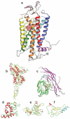
Figure 1
Figure 1
Diversity of the orthosteric ligand-binding domain of various GPCRs. (a) Crystal structure of dark state bovine rhodopsin (PDB ID: 1U19). Rhodopsin is covalently bound to 11-cis-retinal ( pink spheres). Helices are denoted by the following colors: TM-I, blue; TM-II, cyan; TM-III, dark green; TM-IV, lime green; TM-V, yellow; TM-VI, orange; TM-VII, red; and H-8, purple. (b) The ligand-binding domain of the group II metabotropic glutamate receptor (PDB ID: 2E4U). Glutamate (red and green spheres) binds in a crevice formed by the two lobes of the ligand-binding domain. (c) The complex between the ligand-binding domain of the follicle stimulating hormone receptor ( green and yellow) and its ligand, follicle stimulating hormone ( pink and cyan) (PDB ID: 1XWD). (d ) The cysteine-rich domain of murine frizzled 8 (PDB ID: 1IJY). (e) Crystal structure of the ligand-binding domain of the Drosophila methuselah receptor (PDB ID: 1FJR). ( f ) A representative structure of the ligand-binding domain from the NMR ensemble of structures for the corticotropin releasing factor II (CRFR-IIβ) receptor with bound agonist astressin (cyan) (PDB ID: 2JND). All amino terminal ligand-binding domains are oriented such that the putative location of the 7-TM domain would be located below the structure with the exception of the CRFR-IIβ receptor ligand-binding domain for which the orientation is unknown.
Figure 2
Figure 2
Ternary complex model of receptor activity. (a) The classical ternary complex model is shown. Agonists (A) bind free receptor (R) or receptor coupled to the G protein (RG) with the dissociation constants KA and KAG, respectively. The G protein (G) binds free receptor or receptor occupied by agonist (AR) with the dissociation constants KG and KGA, respectively. (b) Curves represent typical patterns revealed in competition studies between a fixed concentration of a radiolabeled antagonist and graded concentrations of an unlabeled agonist. The curves were simulated using parameters obtained from Reference by a two-state multisite model in which a radiolabeled antagonist and an unlabeled agonist compete for sites that are mutually independent and noninterconverting. In the absence of GTP (blue curve), the agonist reveals both a high-affinity state attributed to the receptor coupled to the G protein (RG) and a low-affinity state attributed to free receptor (R). In the presence of GTP (orange curve), the equilibrium appears to shift toward the free receptor state, thereby revealing a single low-affinity state for the agonist. The grey dashed line represents the simulated curve for a receptor exhibiting a single high-affinity state. The ternary complex model is often used to conceptualize the effects shown here; however, this model may not be adequate when applied quantitatively.
Figure 3
Figure 3
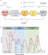
Intermediates of rhodopsin. (a) Rhodopsin is covalently linked to 11-cis-retinal via a protonated Schiff base at Lys296 in transmembrane helix VII. Upon absorption of a photon of light, the chromophore is isomerized to all-trans-retinal. This isomerization leads to the formation of a series of intermediates each exhibiting a distinct absorbance spectrum. The MII state is the active form of the receptor that binds to and activates the G protein. The λmax shown for each intermediate are those from bovine samples (77, 78). The λmax for free 11-cis-retinal and all-trans-retinal are those obtained in ethanol (156). This figure is adapted from References , . (b) Schematic of the reaction coordinate during rhodopsin activation. Isomerization of 11-cis-retinal to all-trans-retinal occurs within femtoseconds with a high quantum yield (Φ). Enthalpies and activation enthalpies for rhodopsin and its intermediates are indicated. Lifetimes of intermediates are shown in parentheses. This figure was adapted from References , .
Figure 4
Figure 4
Structural differences between the dark-state and photoactivated states of rhodopsin. (a, c) Superposition of the three dark-state (PDB ID: 1U19, red; 1GZM, purple; 2I36, cyan) and the early intermediates, bathorhodopsin (PDB ID: 2G87, green) and lumirhodopsin (PDB ID: 2HPY, dark blue). No significant changes are observed except in the region comprising residues 230–243. Panel c is rotated 90° about the x axis. (b, d) Structural superposition of the dark-state (PDB ID:2I36) and the photoactivated state (PDB ID:2I37). Images of crystals for the dark-state and the photoactivated state and their λmax values are shown in the insets. Structural changes observed are limited to cytoplasmic loops II (C-II) and III (C-III) and the cytoplasmic tail. Panel d is rotated 90° about the x axis. The amino terminus and carboxyl terminus are labeled as N and C, respectively.
Figure 5
Figure 5
Model of a rhodopsin oligomer. A model of a rhodopsin oligomer based on distance constraints measured in AFM images of native murine disc membranes is displayed (119) (PDB ID: 1N3M). A top view is shown with helices represented as cylinders and numbered. Two rows of rhodopsin dimers are shown. A pair of rhodopsin dimers packed in the same row is shown on the left. Only one receptor molecule from rhodopsin dimers in the adjacent row on the right is shown. The model is positioned on top of an averaged AFM image of rhodopsin molecules created from raw data presented in Reference .
Figure 6
Figure 6
Oligomeric and dynamic context of receptor activation. (a) The basal state of a dimeric receptor. Thermal fluctuations within the receptor can result in multiple dynamic states of the receptor. Most of the states will be inactive (red ) but a minor population may be constitutively active ( yellow). Different receptor shapes shown represent both dynamic and conformational structural differences. (b) Agonist-bound state of a dimeric receptor. Binding of an agonist (blue square) leads to the activation of the receptor (yellow). An agonist potentially can cause several types of changes in the receptor that result in activity. Binding of an agonist to the receptor can change the distribution or population of receptor states, alter the dimer interface through quaternary changes, and cause cooperativity that affects binding of the next equivalent of an agonist. (c) Binding of both an agonist and a G protein ( green) to the receptor can cause further changes in the structure of the receptor compared with those changes caused by agonist alone. Effects of the agonist and G protein on the dynamics and conformation of the receptor at the tertiary and quaternary levels will define the activation of the receptor.

References

LITERATURE CITED

    1. Fredriksson R, Schioth HB. The repertoire of G-protein-coupled receptors in fully sequenced genomes. Mol. Pharmacol. 2005;67:1414–25. - PubMed
    1. Foord SM, Bonner TI, Neubig RR, Rosser EM, Pin JP, et al. International Union of Pharmacology. XLVI. G protein-coupled receptor list. Pharmacol. Rev. 2005;57:279–88. - PubMed
    1. Filipek S, Stenkamp RE, Teller DC, Palczewski K. G protein-coupled receptor rhodopsin: a prospectus. Annu. Rev. Physiol. 2003;65:851–79. - PMC - PubMed
    1. Filipek S, Teller DC, Palczewski K, Stenkamp R. The crystallographic model of rhodopsin and its use in studies of other G protein-coupled receptors. Annu. Rev. Biophys. Biomol. Struct. 2003;32:375–97. - PMC - PubMed
    1. Ballesteros JA, Shi L, Javitch JA. Structural mimicry in G protein-coupled receptors: implications of the high-resolution structure of rhodopsin for structure-function analysis of rhodopsin-like receptors. Mol. Pharmacol. 2001;60:1–19. - PubMed

RELATED RESOURCES

    1. Kenakin T. Drug efficacy at G protein-coupled receptors. Annu. Rev. Pharmacol. Toxicol. 2002;42:349–79. - PubMed
    1. May LT, Leach K, Sexton PM, Christopoulos A. Allosteric modulation of G protein–coupled receptors. Annu. Rev. Pharmacol. Toxicol. 2007;47:1–51. - PubMed
    1. Palczewski K. G protein-coupled receptor rhodopsin. Annu. Rev. Biochem. 2006;75:743–67. - PMC - PubMed
    1. Smit MJ, Vischer HF, Bakker RA, Jongejan A, Timmerman H, et al. Pharmacogenomic and structural analysis of constitutive G protein–coupled receptor activity. Annu. Rev. Pharmacol. Toxicol. 2007;47:53–87. - PubMed
    1. Spiegel AM, Weinstein LS. Inherited diseases involving G proteins and G protein–coupled receptors. Annu. Rev. Med. 2004;55:27–39. - PubMed

Publication types

LinkOut - more resources