Neuroimaging revolutionizes therapeutic approaches to chronic pain
- PMID: 17848191
- PMCID: PMC2048498
- DOI: 10.1186/1744-8069-3-25
Neuroimaging revolutionizes therapeutic approaches to chronic pain
Abstract
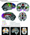
An understanding of how the brain changes in chronic pain or responds to pharmacological or other therapeutic interventions has been significantly changed as a result of developments in neuroimaging of the CNS. These developments have occurred in 3 domains : (1) Anatomical Imaging which has demonstrated changes in brain volume in chronic pain; (2) Functional Imaging (fMRI) that has demonstrated an altered state in the brain in chronic pain conditions including back pain, neuropathic pain, and complex regional pain syndromes. In addition the response of the brain to drugs has provided new insights into how these may modify normal and abnormal circuits (phMRI or pharmacological MRI); (3) Chemical Imaging (Magnetic Resonance Spectroscopy or MRS) has helped our understanding of measures of chemical changes in chronic pain. Taken together these three domains have already changed the way in which we think of pain - it should now be considered an altered brain state in which there may be altered functional connections or systems and a state that has components of degenerative aspects of the CNS.
Figures


Similar articles
-
Central nervous system reorganization in a variety of chronic pain states: a review.PM R. 2011 Dec;3(12):1116-25. doi: 10.1016/j.pmrj.2011.05.018. PM R. 2011. PMID: 22192321 Review.
-
Advances in brain imaging of neuropathic pain.Chin Med J (Engl). 2008 Apr 5;121(7):653-7. Chin Med J (Engl). 2008. PMID: 18466688 Review.
-
Functional imaging of the human trigeminal system: opportunities for new insights into pain processing in health and disease.J Neurobiol. 2004 Oct;61(1):107-25. doi: 10.1002/neu.20085. J Neurobiol. 2004. PMID: 15362156 Review.
-
Brain imaging of neuropathic pain.Neuroimage. 2007;37 Suppl 1:S80-8. doi: 10.1016/j.neuroimage.2007.03.054. Epub 2007 Apr 10. Neuroimage. 2007. PMID: 17512757 Review.
-
Pharmacological modulation of brain activity in a preclinical model of osteoarthritis.Neuroimage. 2013 Jan 1;64:341-55. doi: 10.1016/j.neuroimage.2012.08.084. Epub 2012 Sep 7. Neuroimage. 2013. PMID: 22982372
Cited by
-
Cingulate metabolites during pain and morphine treatment as assessed by magnetic resonance spectroscopy.J Pain Res. 2014 May 19;7:269-76. doi: 10.2147/JPR.S61193. eCollection 2014. J Pain Res. 2014. PMID: 24899823 Free PMC article.
-
Development of a Computerised Adaptive Testing and Equalisation Approaches to Assess Sleep and Quality of Life in Chronic Pain.Eur J Pain. 2025 Oct;29(9):e70108. doi: 10.1002/ejp.70108. Eur J Pain. 2025. PMID: 40844055 Free PMC article.
-
Association between brain N-acetylaspartate levels and sensory and motor dysfunction in patients who have spinal cord injury with spasticity: an observational case-control study.Neural Regen Res. 2023 Mar;18(3):582-586. doi: 10.4103/1673-5374.350216. Neural Regen Res. 2023. PMID: 36018181 Free PMC article.
-
Practical approach to a patient with chronic pain of uncertain etiology in primary care.J Pain Res. 2019 Sep 3;12:2651-2662. doi: 10.2147/JPR.S205570. eCollection 2019. J Pain Res. 2019. PMID: 31564957 Free PMC article.
-
The search for pain biomarkers in the human brain.Brain. 2018 Dec 1;141(12):3290-3307. doi: 10.1093/brain/awy281. Brain. 2018. PMID: 30462175 Free PMC article. Review.
References
-
- Fava M. The role of the serotonergic and noradrenergic neurotransmitter systems in the treatment of psychological and physical symptoms of depression. J Clin Psychiatry. 2003;64 Suppl 13:26–29. - PubMed
MeSH terms
LinkOut - more resources
Full Text Sources
Other Literature Sources
Medical

