. 2007 Sep 12:7:57.
doi: 10.1186/1472-6807-7-57.

Solution structure of the second bromodomain of Brd2 and its specific interaction with acetylated histone tails

Affiliations

Solution structure of the second bromodomain of Brd2 and its specific interaction with acetylated histone tails

Hongda Huang et al. BMC Struct Biol. .

Abstract

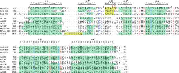
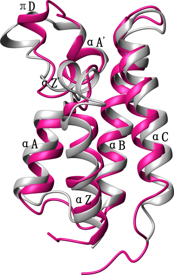
Background: Brd2 is a transcriptional regulator and belongs to BET family, a less characterized novel class of bromodomain-containing proteins. Brd2 contains two tandem bromodomains (BD1 and BD2, 46% sequence identity) in the N-terminus and a conserved motif named ET (extra C-terminal) domain at the C-terminus that is also present in some other bromodomain proteins. The two bromodomains have been shown to bind the acetylated histone H4 and to be responsible for mitotic retention on chromosomes, which is probably a distinctive feature of BET family proteins. Although the crystal structure of Brd2 BD1 is reported, no structure features have been characterized for Brd2 BD2 and its interaction with acetylated histones.

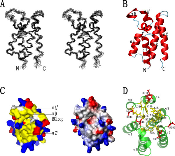
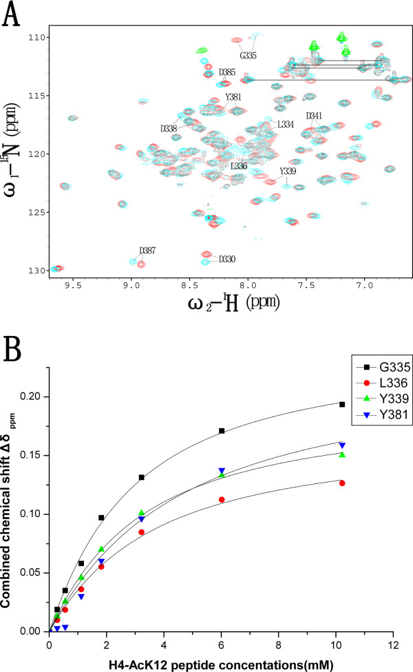
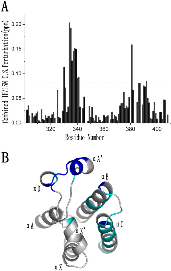
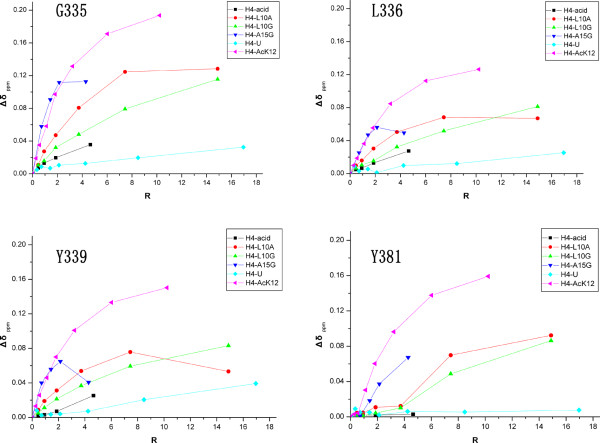
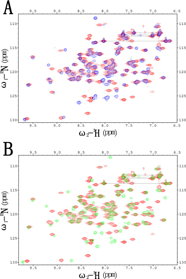
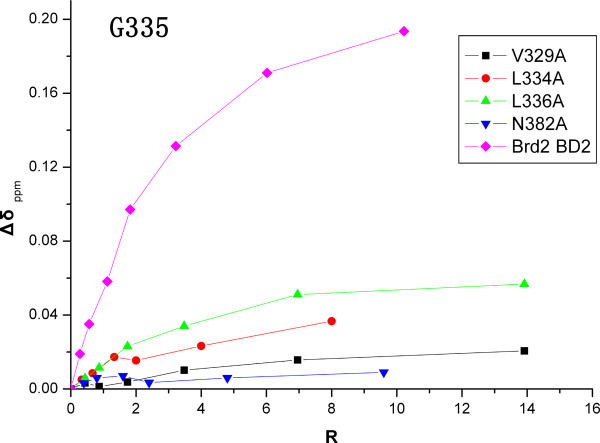
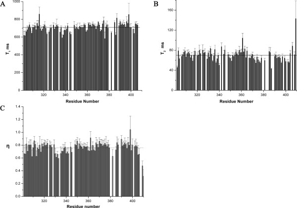
Results: Here we report the solution structure of human Brd2 BD2 determined by NMR. Although the overall fold resembles the bromodomains from other proteins, significant differences can be found in loop regions, especially in the ZA loop in which a two amino acids insertion is involved in an uncommon pi-helix, termed piD. The helix piD forms a portion of the acetyl-lysine binding site, which could be a structural characteristic of Brd2 BD2 and other BET bromodomains. Unlike Brd2 BD1, BD2 is monomeric in solution. With NMR perturbation studies, we have mapped the H4-AcK12 peptide binding interface on Brd2 BD2 and shown that the binding was with low affinity (2.9 mM) and in fast exchange. Using NMR and mutational analysis, we identified several residues important for the Brd2 BD2-H4-AcK12 peptide interaction and probed the potential mechanism for the specific recognition of acetylated histone codes by Brd2 BD2.

Conclusion: Brd2 BD2 is monomeric in solution and dynamically interacts with H4-AcK12. The additional secondary elements in the long ZA loop may be a common characteristic of BET bromodomains. Surrounding the ligand-binding cavity, five aspartate residues form a negatively charged collar that serves as a secondary binding site for H4-AcK12. We suggest that Brd2 BD1 and BD2 may possess distinctive roles and cooperate to regulate Brd2 functions. The structure basis of Brd2 BD2 will help to further characterize the functions of Brd2 and its BET members.


Figures

Figure 1
Figure 1
Structure and molecular contact surfaces of Brd2 BD2. (A) Stereoview of the selected 20 structures of Brd2 BD2, superimposed on backbone atoms (N, Cα and C'). (B) Ribbon representation of the average, energy-minimized structure with the secondary structure elements highlighted. The helix nomenclature follows that of hsP/CAF bromodomain [9]. (C) Contact surface emphasized surface hydrophobic potential (left) and surface electrostatic potential (right) at the acetyl-lysine binding site. Yellow denotes hydrophobic potential; red negative potential; and blue positive potential. (D) A clear view of showing the conserved or type conserved side chains lined the hydrophobic cavity, and denoting the negative-charged collar formed by residues D330, D338, D341, D385 and D387. A, B, C and D were produced with MOLMOL or PyMOL.
Figure 2
Figure 2
Sequence alignment of Brd2 BD2. Sequence alignment of Brd2 BD2 with a selected number of Bromodomains including Brd2 BD1, Brd4 BD2, hsGCN5, scGCN5, hsCBP, hsP/CAF, hsBRG1 and the two components from TAFII250. The sequences were aligned based on the experimentally determined three-dimensional structures of these bromodomains, highlighted in green. The secondary structure of Brd2 BD2 is indicated above the alignment. Residues identical in all sequences are shown in red and residues conserved are coloured in blue and residues corresponding to βZ sheet (hsBRG1) and helix πD are represented in yellow. The two amino acids insertion is indicated by triangle symbols (▼).
Figure 3
Figure 3
Backbone superposition of Brd2 BD2 and BD1 structures. Backbone superposition of the average, energy-minimized structure of Brd2 BD2 (grey) with the crystal structure of Brd2 BD1 (pink) [PDB: 1X0J]. The figure was generated with MOLMOL.
Figure 4
Figure 4
Analysis of the interaction of Brd2 BD2 with H4-AcK12 peptide by NMR. (A) Overlay of the 15N-HSQC spectra of Brd2 BD2 in free of H4-AcK12 peptide (red) and in the presence of H4-AcK12 peptide (cyan). Five of the most perturbed residues (L334, G335, L336, Y339, and Y381) and the five negative-charged aspartate residues (D330, D338, D341, D385 and D387) were denoted. (B) Binding constant was determined by monitoring the combined chemical shift perturbations (Δδppm) of four most perturbed and not severely-overlapped residues (G335, L336, Y339 and Y381) as a function of concentrations of H4-AcK12 peptide.
Figure 5
Figure 5
Binding interface of H4-AcK12 peptide on Brd2 BD2. (A) A histogram view of combined chemical shift perturbations of Brd2 BD2 by addition of H4-AcK12 peptide. The mean value is denoted as a solid line and the mean value plus one standard deviation as a dash line. (B) A ribbon diagram view mapping the binding interface of H4-AcK12 peptide on Brd2 BD2. The residues, whose combined chemical shift changes were more than the mean value plus one standard deviation and above the mean value, are colored in blue and cyan respectively. The figure B was generated in PyMOL.
Figure 6
Figure 6
NMR titrations of Brd2 BD2 with mutated histone H4 tails. Brd2 BD2 was titrated with different H4-AcK12 peptide mutants, including H4-L10A, H4-L10G, H4-A15G, H4-acid and the unacetylated peptide H4-U. Combined chemical shift perturbation was calculated using the equation, Δδppm=(ΔδHN)2+(ΔδNαN)2 MathType@MTEF@5@5@+=feaafiart1ev1aaatCvAUfKttLearuWrP9MDH5MBPbIqV92AaeXatLxBI9gBaebbnrfifHhDYfgasaacH8akY=wiFfYdH8Gipec8Eeeu0xXdbba9frFj0=OqFfea0dXdd9vqai=hGuQ8kuc9pgc9s8qqaq=dirpe0xb9q8qiLsFr0=vr0=vr0dc8meaabaqaciaacaGaaeqabaqabeGadaaakeaacqqHuoariiGacqWF0oazdaWgaaWcbaGaemiCaaNaemiCaaNaemyBa0gabeaakiabg2da9maakaaabaGaeiikaGIaeuiLdqKae8hTdq2aaSbaaSqaaiabdIeaijabd6eaobqabaGccqGGPaqkdaahaaWcbeqaaiabikdaYaaakiabgUcaRiabcIcaOiabfs5aejab=r7aKnaaBaaaleaacqWGobGtaeqaaOGae8xSde2aaSbaaSqaaiabd6eaobqabaGccqGGPaqkdaahaaWcbeqaaiabikdaYaaaaeqaaaaa@489D@, and R = [peptide]/[bromodomain].
Figure 7
Figure 7
Structural integrity analysis of Brd2 BD2 mutants. The integrity of Brd2 BD2 mutants were assessed by their 15N-HSQC spectra. (A) Overlay of the 15N-HSQC spectra of Brd2 BD2 (red) and V329A (blue); (B) Overlay of the 15N-HSQC spectra of Brd2 BD2 (red) and N382A (green).
Figure 8
Figure 8
Plot of Brd2 BD2 mutants titrations with H4-AcK12 peptide. Brd2 BD2 mutants including V329A, L334A, L336A and N382A, were titrated with H4-AcK12 peptide. The doubtless and more significant perturbed residue G335 during titrations was followed.
Figure 9
Figure 9
Plot of the longitudinal and transverse relaxation times and ratio of intensity of NOE. The longitudinal (T1 ms) (A) and transverse relaxation times (T2 ms) (B) and ratio of intensity of NOE (C) are plotted as a function of residue number of Brd2 BD2. Only those residues of which 1H-15N cross-peaks are resolved enough to permit accurate measurements of their intensities are included.

References

    1. Peterson CL, Workman JL. Promoter targeting and chromatin remodeling by the SWI/SNF complex. Curr Opin Genet Dev. 2000;10:187–192. doi: 10.1016/S0959-437X(00)00068-X. - DOI - PubMed
    1. Hassan AH, Neely KE, Vignali M, Reese JC, Workman JL. Promoter targeting of chromatin-modifying complexes. Front Biosci. 2001;6:D1054–1064. doi: 10.2741/Hassan. - DOI - PubMed
    1. Strahl BD, Allis CD. The language of covalent histone modifications. Nature. 2000;403:41–45. doi: 10.1038/47412. - DOI - PubMed
    1. Turner BM. Histone acetylation and an epigenetic code. Bioessays. 2000;22:836–845. doi: 10.1002/1521-1878(200009)22:9<836::AID-BIES9>3.0.CO;2-X. - DOI - PubMed
    1. Turner BM. Cellular memory and the histone code. Cell. 2002;111:285–291. doi: 10.1016/S0092-8674(02)01080-2. - DOI - PubMed

Publication types

MeSH terms