. 2007 Sep 6;14(9):634-44.
doi: 10.1101/lm.607207. Print 2007 Sep.

The central nucleus of the amygdala is essential for acquiring and expressing conditional fear after overtraining

Affiliations

The central nucleus of the amygdala is essential for acquiring and expressing conditional fear after overtraining

Joshua M Zimmerman et al. Learn Mem. .

Abstract

The basolateral complex of the amygdala (BLA) is critical for the acquisition and expression of Pavlovian fear conditioning in rats. Nonetheless, rats with neurotoxic BLA lesions can acquire conditional fear after overtraining (75 trials). The capacity of rats with BLA lesions to acquire fear memory may be mediated by the central nucleus of the amygdala (CEA). To examine this issue, we examined the influence of neurotoxic CEA lesions or reversible inactivation of the CEA on the acquisition and expression of conditional freezing after overtraining in rats. Rats with pretraining CEA lesions (whether alone or in combination with BLA lesions) did not acquire conditional freezing to either the conditioning context or an auditory conditional stimulus after extensive overtraining. Similarly, post-training lesions of the CEA or BLA prevented the expression of overtrained fear. Lastly, muscimol infusions into the CEA prevented both the acquisition and the expression of overtrained fear, demonstrating that the effects of CEA lesions are not likely due to the destruction of en passant axons. These results suggest that the CEA is essential for conditional freezing after Pavlovian fear conditioning. Moreover, overtraining may engage a compensatory fear conditioning circuit involving the CEA in animals with damage to the BLA.


Figures
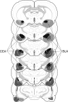
Figure 1.
Figure 1.
Schematic representation of the extent of pretraining NMDA lesions (median lesion) of the CEA (black), BLA (dark gray), and CEA and BLA lesions (light gray) for Experiment 1. Coronal brain section images adapted from Swanson (1992).
Figure 2.
Figure 2.
Conditioned freezing in rats with pretraining amygdala lesions (Experiment 1). (A) Mean percentage of freezing (±SEM) during the 75 trial training session (data are displayed with a 30-sec pretrial period followed by 15 bins consisting of 5 trials each). Freezing was quantified before the first conditioning trial (Pre) and during the 1-min period after each conditioning trial; these values were averaged in 5-trial blocks. The inset shows the mean percentage of freezing (±SEM) during the preperiod through the first two training blocks expanded to show minutes 1–10. (B) Mean percentage of freezing (±SEM) to contextual (8-min context extinction test) cues 24 h after training. (C) Mean percentage of freezing (±SEM) to the auditory CS in a novel context 48 h after training. The auditory CS was initiated 2 min after rats were placed in the chambers (horizontal bar indicates the CS). Data are shown for rats with lesions of the BLA (solid circles), CEA (open circles), or CEA + BLA (open squares); SH rats (solid squares).
Figure 3.
Figure 3.
Schematic representation of the extent of post-training NMDA lesions (median lesion) of the CEA (black), BLA (dark gray), and CEA + BLA lesions (light gray) for Experiment 2. Coronal brain section images adapted from Swanson (1992).
Figure 4.
Figure 4.
Representative slices stained with thionin and AuCl. Slices shown from rats that received lesions of the CEA + BLA (upper right), CEA (lower left), or BLA (lower right); SH rats (upper left). Both the thionin- and the AuCl-stained slices for each group are taken from the same representative animal at approximately the same anterior–posterior level with a magnification of the amygdala shown to the immediate right.
Figure 5.
Figure 5.
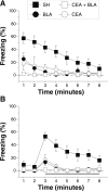
Conditioned freezing in rats with post-training amygdala lesions (Experiment 2). (A) Mean percentage of freezing (±SEM) to contextual (8-min context extinction test) cues after at least 7 d of recovery from post-training surgery. (B) Mean percentage of freezing (±SEM) to the auditory CS in a novel context 24 h after contextual testing. The auditory CS was initiated 2 min after rats were placed in the chambers (horizontal bar indicates the CS). Data are shown for rats with lesions of the BLA (solid circles), CEA (open circles), or CEA + BLA (open squares); SH rats (solid squares).
Figure 6.
Figure 6.
Schematic representation of the extent of pretraining NMDA lesions (median lesion) of the BLA (dark gray) and the locations of included cannula placements for the infusion of muscimol (circles) or 0.9% sterile saline (squares) in the CEA (Experiment 3). A magnification of the amygdala is shown in the insets adjacent to the coronal brain sections. Coronal brain section images adapted from Swanson (1992).
Figure 7.
Figure 7.
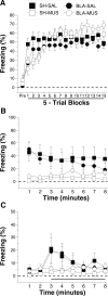
Conditioned freezing in rats with pretraining BLA lesions and temporary inactivation of the CEA during training (Experiment 3). (A) Mean percentage of freezing (±SEM) during the 75 trial training session (data are displayed with a 3-min pretrial period followed by 15 bins consisting of 5 trials each) after infusion of 0.9% saline or muscimol into the CEA. Freezing was quantified before the first conditioning trial (Pre) and during the 1-min period after each conditioning trial; these values were averaged in 5-trial blocks. (B) Mean percentage of freezing (±SEM) to contextual (8-min context extinction test) cues 24 h after training. (C) Mean percentage of freezing (±SEM) to the auditory CS in a novel context 48 h after training. The auditory CS was initiated 2 min after rats were placed in the chambers (horizontal bar indicates the CS). Data are shown for rats with pretraining sham surgeries receiving saline in the CEA before training (SH-SAL: solid squares), pretraining sham surgeries receiving muscimol in the CEA before training (SH-MUS: open squares), pretraining NMDA lesions of the BLA receiving saline in the CEA before training (BLA-SAL: solid circles), or pretraining NMDA lesions of the BLA receiving muscimol in the CEA before training (BLA-MUS: open circles).
Figure 8.
Figure 8.
Shock reactivity in rats with pretraining BLA lesions and temporary inactivation of the CEA before training (Experiment 3). Mean percentage of activity (±SEM) before the first conditioning trial (Pre) and during the first 2-sec shock (Shock) during the conditioning session. Data are shown for rats with pretraining sham surgeries receiving saline in the CEA before training (SH-SAL: solid squares), pretraining sham surgeries receiving muscimol in the CEA before training (SH-MUS: open squares), pretraining NMDA lesions of the BLA receiving saline in the CEA before training (BLA-SAL: solid circles), or pretraining NMDA lesions of the BLA receiving muscimol in the CEA before training (BLA-MUS: open circles).
Figure 9.
Figure 9.
Schematic representation of the extent of pretraining NMDA lesions (median lesion) of the BLA (dark gray) and the locations of included cannula placements for the infusion of muscimol (circles) or 0.9% sterile saline (squares) in the CEA (Experiment 4). A magnification of the amygdala is shown adjacent to the coronal brain sections. Coronal brain section images adapted from Swanson (1992).
Figure 10.
Figure 10.
Conditioned freezing in rats with pretraining BLA lesions and temporary inactivation of the CEA during testing (Experiment 4). (A) Mean percentage of freezing (±SEM) during the 75 trial training session (data are displayed with a 3-min pretrial period followed by 15 bins consisting of 5 trials each). Freezing was quantified before the first conditioning trial (Pre) and during the 1-min period after each conditioning trial; these values were averaged in 5-trial blocks. (B) Mean percentage of freezing (±SEM) to contextual (8-min context extinction test) cues after infusion of 0.9% sterile saline or muscimol into the CEA. (C) Mean percentage of freezing (±SEM) to the auditory CS in a novel context 48 h after training after infusion of 0.9% sterile saline or muscimol into the CEA. The auditory CS was initiated 2 min after rats were placed in the chambers (horizontal bar indicates the CS). Data are shown for rats with pretraining sham surgeries receiving saline in the CEA before testing (SH-SAL: solid squares), pretraining sham surgeries receiving muscimol in the CEA before testing (SH-MUS: open squares), pretraining NMDA lesions of the BLA receiving saline in the CEA before testing (BLA-SAL: solid circles), or pretraining NMDA lesions of the BLA receiving muscimol in the CEA before testing (BLA-MUS: open circles).

Similar articles

Cited by

References

    1. Anglada-Figueroa D., Quirk G.J. Lesions of the basal amygdala block expression of conditioned fear but not extinction. J. Neurosci. 2005;25:9680–9685. - PMC - PubMed
    1. Balleine B.W., Killcross S. Parallel incentive processing: An integrated view of amygdala function. Trends Neurosci. 2006;29:272–279. - PubMed
    1. Bevins R.A., McPhee J.E., Rauhut A.S., Ayres J.J. Converging evidence for one-trial context fear conditioning with an immediate shock: Importance of shock potency. J. Exp. Psychol. Anim. Behav. Process. 1997;23:312–324. - PubMed
    1. Cahill L., Vazdarjanova A., Setlow B. The basolateral amygdala complex is involved with, but is not necessary for, rapid acquisition of Pavlovian 'fear conditioning.'. Eur. J. Neurosci. 2000;12:3044–3050. - PubMed
    1. Campeau S., Davis M. Involvement of the central nucleus and basolateral complex of the amygdala in fear conditioning measured with fear-potentiated startle in rats trained concurrently with auditory and visual conditioned stimuli. J. Neurosci. 1995;15:2301–2311. - PMC - PubMed

Publication types

MeSH terms