. 2007 Sep 18;104(38):15000-5.
doi: 10.1073/pnas.0703738104. Epub 2007 Sep 11.

Proteasomal selection of multiprotein complexes recruited by LIM homeodomain transcription factors

Affiliations

Proteasomal selection of multiprotein complexes recruited by LIM homeodomain transcription factors

Cenap Güngör et al. Proc Natl Acad Sci U S A. .

Erratum in

  • Proc Natl Acad Sci U S A. 2007 Oct 23;104(43):17239

Abstract

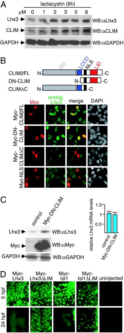
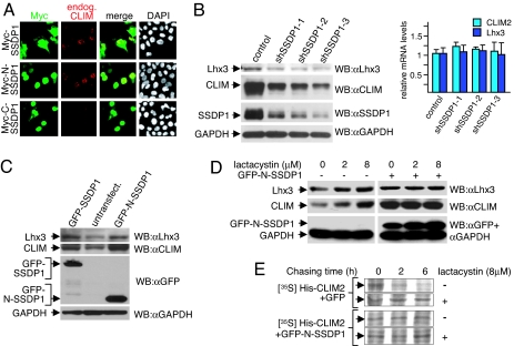
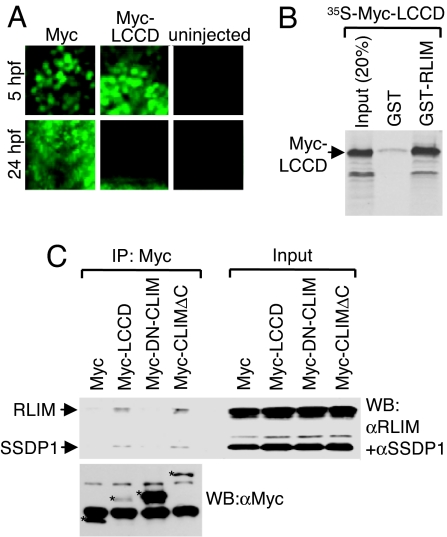
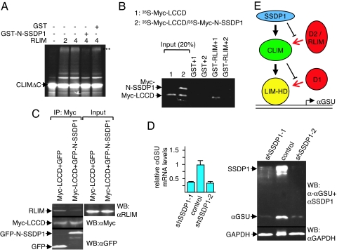
Complexes composed of multiple proteins regulate most cellular functions. However, our knowledge about the molecular mechanisms governing the assembly and dynamics of these complexes in cells remains limited. The in vivo activity of LIM homeodomain (LIM-HD) proteins, a class of transcription factors that regulates neuronal development, depends on the high-affinity association of their LIM domains with cofactor of LIM homeodomain proteins (LIM-HDs) (CLIM, also known as Ldb or NLI). CLIM cofactors recruit single-stranded DNA-binding protein 1 (SSDP1, also known as SSBP3), and this interaction is important for the activation of the LIM-HD/CLIM protein complex in vivo. Here, we identify a cascade of specific protein interactions that protect LIM-HD multiprotein complexes from proteasomal degradation. In this cascade, CLIM stabilizes LIM-HDs, and SSDP1 stabilizes CLIM. Furthermore, we show that stabilizing cofactors prevent binding of ubiquitin ligases to multiple protein interaction domains in LIM-HD recruited protein complexes. Together, our results indicate a combinatorial code that selects specific multiprotein complexes via proteasomal degradation in cells with broad implications for the assembly and specificity of multiprotein complexes.


Conflict of interest statement

The authors declare no conflict of interest.

Figures

Fig. 1.
Fig. 1.
CLIM protects endogenous Lhx3 from proteasomal degradation in αT3 cells. (A) Cellular levels of Lhx3 and CLIM are regulated by the proteasome. Shown are Western blots of protein extracts that were treated for 6 h with increasing amounts of proteasome inhibitor lactacystin. (B) (Upper) CLIM deletion constructs consisting of CLIM2 full length (CLIM2FL; 1–373), dominant-negative CLIM (DN-CLIM; 245–341), and a C-terminal deletion (CLIMΔC; 1–277). DD, dimerization domain; LCCD, Ldb1/Chip conserved domain; NLS, nuclear localization signal; LID, LIM interaction domain. (Lower) Transfection of αT3 cells with Myc-tagged CLIM expression constructs. Cells are costained with specific Myc (red) and Lhx3 (green) antisera. Note the higher levels of endogenous Lhx3 in cells transfected with Myc-CLIM2FL and Myc-DN-CLIM. (C) (Left) Western blot of protein extracts of cells transfected with Myc-DN-CLIM or the empty vector as control. The same blot was probed with antisera against Lhx3, Myc and GAPDH. Note the higher levels of endogenous Lhx3 in cells transfected with Myc-DN-CLIM. (Right) No change in Lhx3 mRNA levels of the same transfected cells as measured by real-time RT-PCR (n = 3; values are mean ± SE). (D) The LIM domain mediates instability of LIM-HD proteins. mRNA encoding LIM-HD proteins Lhx3 and Isl1 with or without LIM domains were injected in one- to two-cell-stage zebrafish embryos. Embryos were fixed at 5 and 24 hpf and stained with a monoclonal Myc antibody. At 24 hpf, the focus is on trunk somites. Note that little or no Myc staining is detected in Myc-Lhx3 and Myc-Isl1-injected embryos, whereas ΔLIM proteins are readily detectable.
Fig. 2.
Fig. 2.
A cascade of protein interactions protects LIM-HD complexes from proteasomal degradation. (A) αT3 cells were transfected with Myc-SSDP1 expression constructs. Cells are costained with Myc (red) and CLIM (green) antibodies. Note the higher levels of endogenous CLIM in nuclei of cells transfected with Myc-SSDP1 and Myc-N-SSDP1 (1–92) containing the CLIM-interaction domain, but not Myc-C-SSDP1 (90–361). (B) Knock-down of SSDP1 results in decreased levels of endogenous CLIM and Lhx3. SSDP1 levels were knocked-down in αT3 cells via retroviral infection of three independent mouse shRNAs directed against different regions on SSDP1 or the empty retroviral vector as control. Note that knocking down endogenous SSDP1 levels in αT3 cells leads to a decrease in endogenous CLIM and Lhx3 protein levels (Left), whereas mRNA levels remain unchanged as measured by qRT-PCR (Right), (n = 3; values are mean ± SE). (C) Western blot of protein extracts from αT3 cells transfected with GFP-SSDP1 and GFP-N-SSDP1 expression constructs. GFP-positive cells were isolated via FACS before extract preparation. GFP-negative cells from the same sorting were used as negative control. The same blot was probed with antisera against Lhx3, CLIM, GFP, and GAPDH. Note the higher levels of endogenous CLIM and Lhx3 in cells overexpressing GFP-SSDP1 or GFP-N-SSDP1. (D) SSDP1 protects the Lhx3-CLIM complex from proteasomal degradation. αT3 cells were transfected with GFP-N-SSDP1 expression construct, and GFP-positive cells were isolated via FACS. Both GFP-positive and GFP-negative cells of the same sorting were cultured overnight. Cells were then treated with indicated concentrations of lactacystin for 6 h before harvesting for Western blotting. The same blot was probed with antisera against Lhx3, CLIM, and GFP/GAPDH. Note that Lhx3 and CLIM protein levels in GFP-N-SSDP1-expressing cells are no longer sensitive to lactacystin. (E) SSDP1 increases the half-life of CLIM. His-tagged CLIM2 was coexpressed in HEK293T cells with GFP or GFP-N-SSDP1 and total cellular proteins were labeled with [35S]methionine for 1 h, followed by washings and chasing in normal medium for indicated time periods in the presence or absence of lactacystin.
Fig. 3.
Fig. 3.
The LCCD region interacts with SSDP1 and RLIM and mediates instability. (A) The LCCD domain functions as an instability domain. mRNA encoding Myc-LCCD was injected in zebrafish embryos. Embryos were fixed at 5 and 24 hpf and stained with a monoclonal Myc antibody. Note that little or no Myc staining is detected in 24 hpf embryos of Myc-LCCD-injected animals, whereas Myc-NLS control proteins are readily detectable. (B) The LCCD region interacts directly with RLIM in vitro. Shown is GST pull-down using GST-RLIM and [35S]Myc-LCCD. (C) The LCCD interacts with RLIM and SSDP1 in cells. Co-IP of SSDP1 and RLIM from αT3 cells transfected with Myc-LCCD. (Upper) Western blot of Myc IPs (Left) and 100% input control (Right) using antibodies directed against RLIM and SSDP1 (10% gel). (Lower) To visualize Myc-proteins, an aliquot of the same Myc-IPs was run on a 15% gel in parallel. Myc proteins are indicated by asterisks. Lower Myc-LCCD and Myc-CLIMΔC protein levels probably reflect their instability in cells. Bands not marked with asterisks are unspecific.
Fig. 4.
Fig. 4.
SSDP1 inhibits binding of RLIM to CLIM and regulates Lhx3 target gene expression. (A) In vitro ubiquitination experiment using 35S-labeled CLIMΔC, RLIM as E3, and UbcH5 as E2 enzyme in the presence of GST-N-SSDP1 or GST alone. Arrows point at unmodified CLIM proteins. Polyubiquitinated proteins are indicated by three asterisks (***). Note the partial inhibition of CLIMΔC ubiquitination by N-SSDP1. (B) N-SSDP1 inhibits binding of RLIM to the LCCD. Shown is the GST pull-down using GST-RLIM and 35S-Myc-LCCD (1) or Myc-LCCD cotranslated with Myc-N-SSDP1 (35S-Myc-LCCD/35S-Myc-N-SSDP1) (2). (C) Co-IP of endogenous RLIM in cells cotransfected with Myc-LCCD and GFP-N-SSDP1. Note the decreased RLIM precipitation in the presence of N-SSDP1. (D) Down-regulation of SSDP1 results in lower levels of αGSU mRNA and protein. (Left) SSDP1 levels were knocked-down in αT3 cells via retroviral infection of mouse shRNAs (shSSDP1-1, -2) or the empty retroviral vector. mRNA encoding αGSU was measured by qRT-PCR (n = 3; values are mean ± SE). (Right) Western blot of the same shRNA-treated cells. Note that knocking down endogenous SSDP1 levels leads to a significant decrease in endogenous αGSU levels at the mRNA and protein levels. (E) Shown is a model of a cascade of protein interactions that protects LIM-HDs from proteasomal degradation. Via binding to LIM domains, a destabilizing enzyme (D1, red) targets LIM-HDs (yellow) for degradation. In the presence of CLIM (green), the binding of D1 to LIM domains is inhibited, resulting in stabilization of LIM-HDs. CLIM is targeted by another destabilizing enzyme(s) (D2/RLIM) for ubiquitination/degradation. The presence of SSDP1 (blue) prevents binding of D2/RLIM to CLIM thereby protecting the LIM-HD/CLIM protein complex.

Similar articles

Cited by

References

    1. Bach I. Mech Dev. 2000;91:5–17. - PubMed
    1. Hobert O, Westphal H. Trends Genet. 2000;16:75–83. - PubMed
    1. Matthews JM, Visvader JE. EMBO Rep. 2003;4:1132–1137. - PMC - PubMed
    1. Chen L, Segal D, Hukriede NA, Podtelejnikov AV, Bayarsaihan D, Kennison JA, Ogryzko VV, Dawid IB, Westphal H. Proc Natl Acad Sci USA. 2002;99:14320–14325. - PMC - PubMed
    1. van Meyel DJ, Thomas JB, Agulnick AD. Development (Cambridge, UK) 2003;130:1915–1925. - PubMed

Publication types

MeSH terms

Substances