. 2007 Sep 18;104(38):14905-10.
doi: 10.1073/pnas.0700717104. Epub 2007 Sep 11.

A structural gap in Dpo4 supports mutagenic bypass of a major benzo[a]pyrene dG adduct in DNA through template misalignment

Affiliations

A structural gap in Dpo4 supports mutagenic bypass of a major benzo[a]pyrene dG adduct in DNA through template misalignment

Jacob Bauer et al. Proc Natl Acad Sci U S A. .

Erratum in

  • Proc Natl Acad Sci U S A. 2007 Oct 23;104(43):17240

Abstract

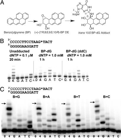
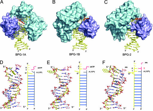
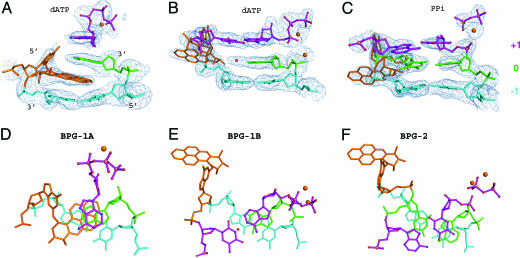
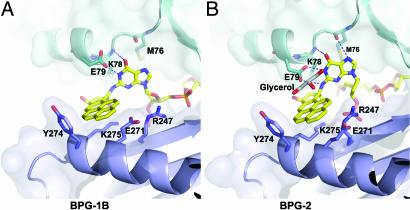
Erroneous replication of lesions in DNA by DNA polymerases leads to elevated mutagenesis. To understand the molecular basis of DNA damage-induced mutagenesis, we have determined the x-ray structures of the Y-family polymerase, Dpo4, in complex with a DNA substrate containing a bulky DNA lesion and incoming nucleotides. The DNA lesion is derived from an environmentally widespread carcinogenic polycyclic aromatic hydrocarbon, benzo[a]pyrene (BP). The potent carcinogen BP is metabolized to diol epoxides that form covalent adducts with cellular DNA. In the present study, the major BP diol epoxide adduct in DNA, BP-N(2)-deoxyguanosine (BP-dG), was placed at a template-primer junction. Three ternary complexes reveal replication blockage, extension past a mismatched lesion, and a -1 frameshift mutation. In the productive structures, the bulky adduct is flipped/looped out of the DNA helix into a structural gap between the little finger and core domains. Sequestering of the hydrophobic BP adduct in this new substrate-binding site permits the DNA to exhibit normal geometry for primer extension. Extrusion of the lesion by template misalignment allows the base 5' to the adduct to serve as the template, resulting in a -1 frameshift. Subsequent strand realignment produces a mismatched base opposite the lesion. These structural observations, in combination with replication and mutagenesis data, suggest a model in which the additional substrate-binding site stabilizes the extrahelical nucleotide for lesion bypass and generation of base substitutions and -1 frameshift mutations.


Conflict of interest statement

The authors declare no conflict of interest.

Figures

Fig. 1.
Fig. 1.
Formation of a benzo[a]pyrene diol epoxide adduct (BP–dG) and its effect on DNA replication. (A) Structures of BP, its (+)-(7R,8S,9S,10R) diol epoxide (DE) metabolite and the trans 10S BP–dG adduct in DNA. The α′, β′, and χ torsion angles are labeled. (B) Incorporation of nucleotides opposite the BP–dG adduct in the template in comparison with incorporation opposite an unmodified dG residue. The BP–dG adduct is indicated as G*. Assays were performed with the dNTP concentrations and the times indicated. Lanes 0, 4, G, A, T, and C correspond to no dNTP, all four dNTPs, and each single dNTP, respectively. ddC (Right) indicates the 3′ end of the template strand is dideoxy-C, which is used in crystallization to prevent nucleotide addition at the blunt end (21). (C) Incorporation of dNTP opposite the BP–dG adduct in four sequence contexts by Dpo4. The arrows indicate the expected size of the full-length products. Reaction time was 1 hour with 100 μM dNTPs.
Fig. 2.
Fig. 2.
Structures of BPG-1A, BPG-1B and BPG-2. (A–C) Dpo4 is represented as a molecular surface with the polymerase core in cyan and the LF domain in purple; DNA and nucleotide are shown as sticks, and BP–dG is highlighted in orange. BPG-1B and BPG-2 in B and C are rotated 180° relative to BPG-1A in A around the DNA helix axis, to show the extrahelical BP–dG in the gap between the core and LF domains. (D–F) The DNA conformations corresponding to (A–C) as stick models, all with the same orientations as in A. The primer strands are in gray, and incoming dATP is in pink. The single-stranded portion of the template DNA is not shown. Figs. 2, 3, and 4 were generated by using PYMOL (46).
Fig. 3.
Fig. 3.
The BP–dG adduct and its surrounding base pairs in the active site. The base pairs are colored according to their position relative to the lesion site: The lesion site (0) is green, +1 (above) is purple, and −1 is light cyan. The adducted nucleotide itself is orange. The DNA and dATP are shown as ball-and-stick models with Ca2+ ions and water molecules shown as orange and red spheres. (A–C) Side views of the base pairs with the simulated annealing omit map contoured at 2.5σ at 2.5 Å and 2.25 Å resolution, respectively. (D–F) Views rotated 90° relative to the corresponding image in A–C. The oxygen, nitrogen, and phosphorus atoms are colored red, blue, and pink, respectively, for the top layer.
Fig. 4.
Fig. 4.
Close-up views of BP–dG in the structure gap between the core and the LF domains. (A) BPG-1B. (B) BPG-2. The protein is in ribbon models covered by a transparent molecular surface. The key residues interacting with the adduct G* are shown as stick models. The BP ring system is in van der Waals contact with the LF domain (purple); the adducted G base interacts with the core domain (cyan). The glycerol molecule is in gray.

References

    1. Friedberg EC, Wagner R, Radman M. Science. 2002;296:1627–1630. - PubMed
    1. Goodman MF. Annu Rev Biochem. 2002;71:17–50. - PubMed
    1. Lehmann AR. Mutat Res. 2002;509:23–34. - PubMed
    1. Committee on the Biological Effects of Atmospheric Pollutants. Washington DC: Natl Acad Sci; 1972.
    1. Phillips DH. Mutat Res. 1999;443:139–147. - PubMed

Publication types

MeSH terms

LinkOut - more resources