. 2007 Nov 9;282(45):32974-82.
doi: 10.1074/jbc.M706529200. Epub 2007 Sep 11.

Restricted active site docking by enzyme-bound substrate enforces the ordered cleavage of prothrombin by prothrombinase

Affiliations

Restricted active site docking by enzyme-bound substrate enforces the ordered cleavage of prothrombin by prothrombinase

Ayse Hacisalihoglu et al. J Biol Chem. .

Abstract

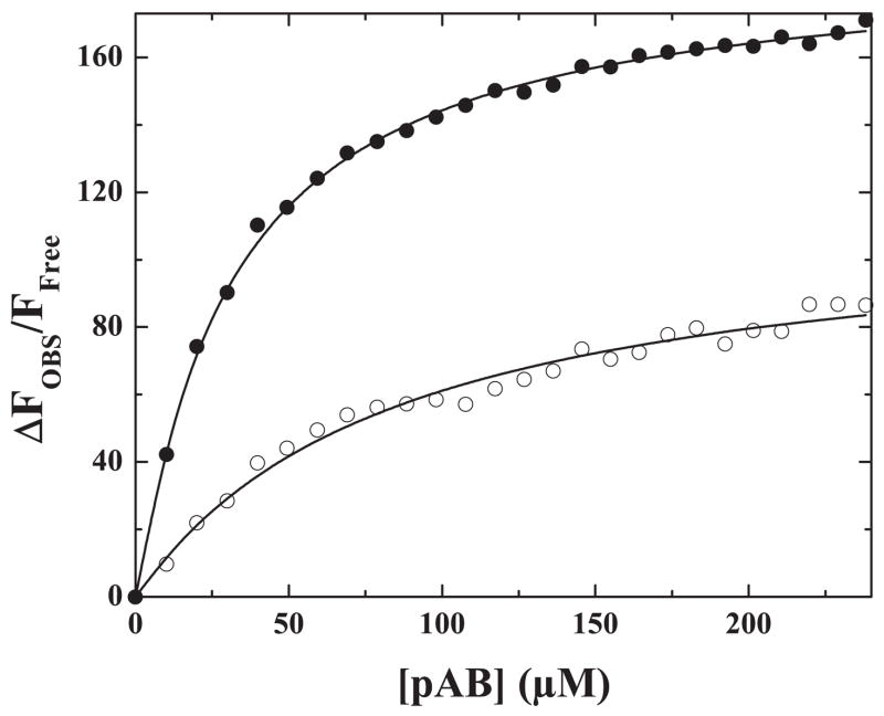
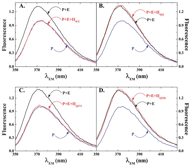
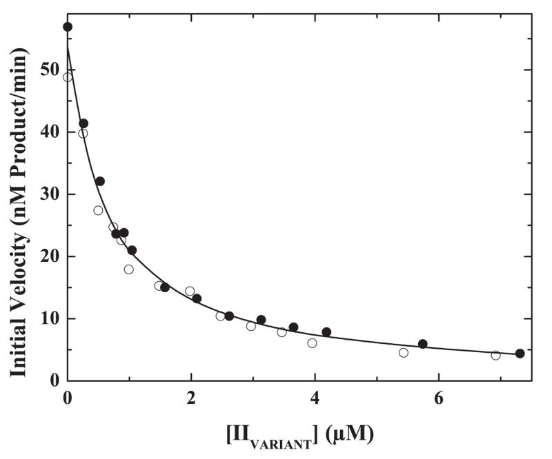
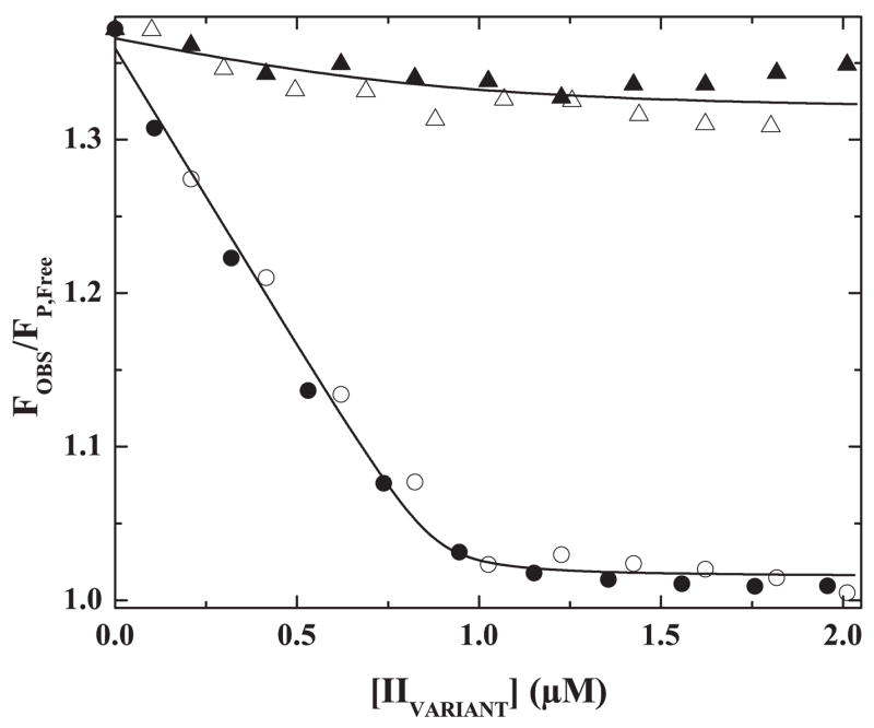
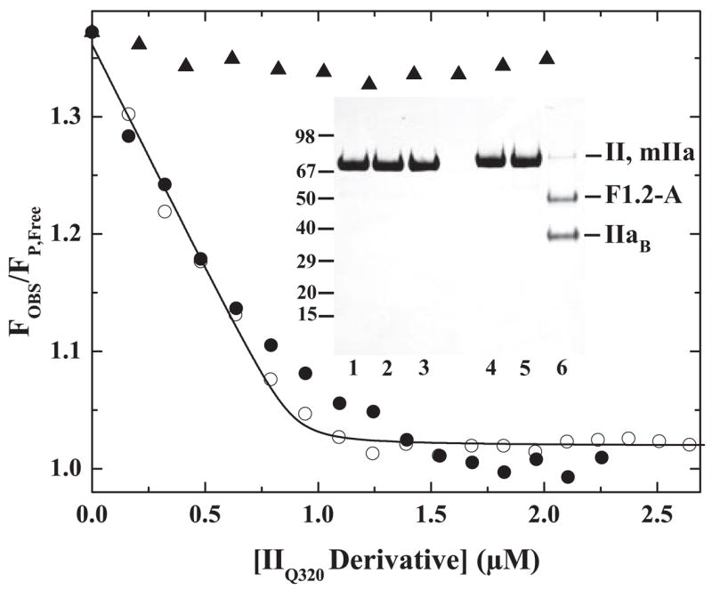
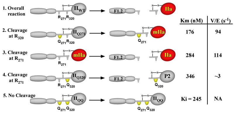
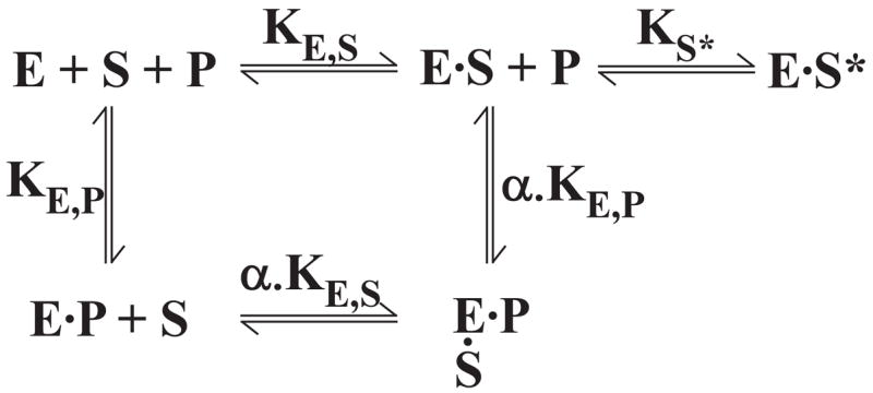
The preferred pathway for prothrombin activation by prothrombinase involves initial cleavage at Arg(320) to produce meizothrombin, which is then cleaved at Arg(271) to liberate thrombin. Exosite binding drives substrate affinity and is independent of the bond being cleaved. The pathway for cleavage is determined by large differences in V(max) for cleavage at the two sites within intact prothrombin. By fluorescence binding studies in the absence of catalysis, we have assessed the ability of the individual cleavage sites to engage the active site of Xa within prothrombinase at equilibrium. Using a panel of recombinant cleavage site mutants, we show that in intact prothrombin, the Arg(320) site effectively engages the active site in a 1:1 interaction between substrate and enzyme. In contrast, the Arg(271) site binds to the active site poorly in an interaction that is approximately 600-fold weaker. Perceived substrate affinity is independent of active site engagement by either cleavage site. We further show that prior cleavage at the 320 site or the stabilization of the uncleaved zymogen in a proteinase-like state facilitates efficient docking of Arg(271) at the active site of prothrombinase. Therefore, we establish direct relationships between docking of either cleavage site at the active site of the catalyst, the V(max) for cleavage at that site, substrate conformation, and the resulting pathway for prothrombin cleavage. Exosite tethering of the substrate in either the zymogen or proteinase conformation dictates which cleavage site can engage the active site of the catalyst and enforces the sequential cleavage of prothrombin by prothrombinase.


Figures

FIGURE 1
FIGURE 1. Binding of pAB to Xa and prothrombinase
Fluorescence titrations were performed as described under “Experimental Procedures” by the addition of increasing concentrations of pAB to 1 μM Xa (○) or to 1 μM prothrombinase (1 μM Xa, 1.2 μM Va, 200 μM PCPS) (●). Fluorescence was corrected, normalized, and analyzed as described. The lines are drawn using the fitted parameters listed in Table 1.
FIGURE 2
FIGURE 2. Displacement of pAB from prothrombinase
Technical fluorescence emission spectra (λEX = 320 nm) were obtained using reaction mixtures containing 25 μM pAB, 200 μM PCPS, 1.2 μM Va (P), the reaction mixture in P but also containing 1 μM XaS195A (P + E), and the reaction mixture in P + E also containing 1.4 μM IIWT (panel A, P + E + IIWT), 1.4 μM IIQQ (panel B, p + E + IIQQ), 1.4 μM IIQ271 (panel C, P + E + IIQ271) or 1.4 μM IIQ320 (panel D, P + E + IIQ320). Each spectrum, collected by averaging three scans, was corrected for scattering and normalized by setting the peak fluorescence of reaction P to 1.
FIGURE 3
FIGURE 3. Inhibition of IIQ271 cleavage
Initial velocities for the cleavage of 300 nM IIQ271 by 0.02 nM prothrombinase (0.02 nM Xa, 31 nM Va, 27.5 μM PCPSLUV) were determined in the presence of increasing concentrations of IIQ320 (●) or IIQQ (○). The line is drawn following simultaneous analysis of both data sets with the rate expression for classical competitive inhibition using an assumed value of V/E = 94 s−1 (Scheme I) and fitted values for Km = 231 ± 11 nM and Ki = 336 ± 23 nM.
FIGURE 4
FIGURE 4. Active site docking by prothrombin and its variants
Fluorescence intensity normalized to that of free pAB was measured in reaction mixtures containing 25 μM pAB, 1 μM prothrombinase (1 μM XaS195A, 1.2 μM Va, 200 μM PCPS), and 10 μM FPR-CH2Cl with increasing concentrations of IIWT (●), IIQ271 (○), IIQ320 (▲), or IIQQ (△). The line drawn through the IIWT and IIQ271 data sets was obtained by analysis according to Scheme II with the indicated assumptions and the fitted terms ΔFMAXFFREE = 31.3 ± 1.4, 0.88 ± 0.03 mol of S/mol of E at saturation and Ks* ≤ 0.017. The line drawn through the IIQQ and IIQ320 data sets corresponds to the fitted terms ΔFMAXFFREE = 34.1 ± 2.3 and Ks* ≥12.3 assuming 0.88 mol of S/mol of E at saturation.
FIGURE 5
FIGURE 5
Active site engagement by Arg271 upon proteinase formation. Normalized fluorescence intensity of pAB was measured in reaction mixtures containing 25 μM pAB, 1 μM prothrombinase (1 μM XaS195A, 1.2 μM Va, 200 μM PCPS), and 10 μM FPR-CH2Cl with increasing concentrations of FPR-II320 (●) or mIIa produced from IIQ320 (○). Data obtained with increasing concentrations of IIQ320 (▲) are provided for reference. The line drawn by analysis of data obtained with mIIa according to Scheme II and the fitted terms ΔFMAXFFREE = 30.8 ± 1.1, 0.89 ± 0.02 mol of S/mol of E at saturation, and Ks* ≤ 0.018. Inset, SDS-PAGE analysis of IIQ320 (lanes 1 and 4), FPR-IIQ320 (lanes 2 and 5), and mIIa produced from IIQ320 (lanes 3 and 6) either before (lanes 1–3) or after (lanes 4 – 6) disulfide bond reduction. Approximately 5 μg of protein was loaded per lane.
SCHEME I
SCHEME I. Cleavage of prothrombin and variants
Prothrombinase converts prothrombin (IIWT) to thrombin (IIa) and the activation peptide F1.2 by catalyzing cleavage at Arg271 and Arg320 (reaction 1). The individual reactions have been studied using recombinant derivatives (IIQ271, IIQ320) or the proteolytic intermediate mIIa with the indicated representative steady state kinetic constants (12). The overall reaction can be accounted for by the essentially ordered cleavage of prothrombin at Arg320 to form mIIa (reaction 2) followed by cleavage at Arg271 (reaction 3). Cleavage of bonds in the opposite order is not readily detected because of the low Vmax for cleavage at Arg271 in IIQ320 to yield prethrombin 2 (P2) and F1.2 (reaction 4). Although IIQQ is not cleaved by prothrombinase (reaction 5), it acts as a competitive inhibitor for the cleavage of the other substrates.
SCHEME II
SCHEME II. Equilibria for the binding of active site-directed probe and substrate to prothrombinase
The protein substrate (S) binds to prothrombinase (E) through an initial exosite binding step determined by KE,S followed by active site engagement to yield ES*. The equilibrium constant for active site docking is given by Ks*. This dimensionless unimolecular equilibrium constant is defined as Ks* = [ES]/[ES*]. The binding of pAB (P) to the active site is determined by KE,P. Based on previous kinetic studies, α is assumed to be indistinguishable from 1 (15).

References

    1. Jackson CM, Nemerson Y. Annu Rev Biochem. 1980;49:765–811. - PubMed
    1. Mann KG, Jenny RJ, Krishnaswamy S. Annu Rev Biochem. 1988;57:915–956. - PubMed
    1. Mann KG, Nesheim ME, Church WR, Haley P, Krishnaswamy S. Blood. 1990;76:1–16. - PubMed
    1. Camire RM, Pollack ES. In: Hemostasis and Thrombosis, Basic Principles and Clinical Practice. Colman RW, Marder VJ, Clowes AJ, George JN, Goldhaber SZ, editors. Lippincott Williams & Wilkins; Philadelphia: 2006. pp. 59–89.
    1. Jenny NS, Lundblad RL, Mann KG. In: Hemostasis and Thrombosis, Basic Principles and Clinical Practice. Colman RW, Marder VJ, Clowes AJ, George JN, Goldhaber SZ, editors. Lippincott Williams & Wilkins; Philadelphia: 2006. pp. 193–213.

Publication types

LinkOut - more resources