. 2007 Nov 9;282(45):33168-80.
doi: 10.1074/jbc.M703736200. Epub 2007 Sep 11.

Activity-dependent regulation of h channel distribution in hippocampal CA1 pyramidal neurons

Affiliations

Activity-dependent regulation of h channel distribution in hippocampal CA1 pyramidal neurons

Minyoung Shin et al. J Biol Chem. .

Abstract

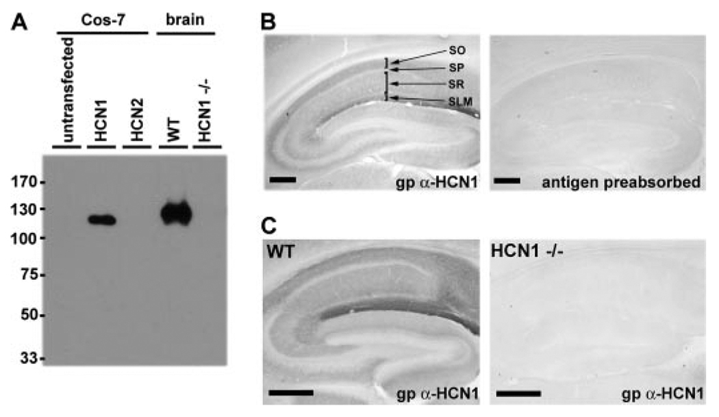
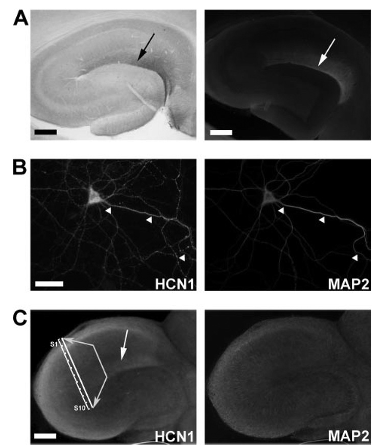
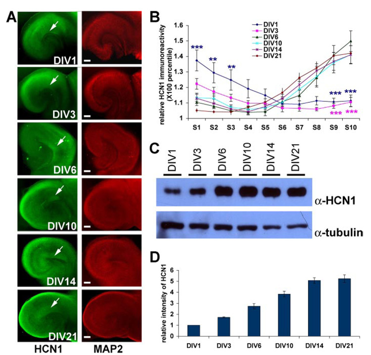
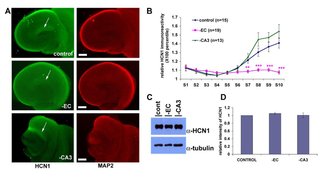
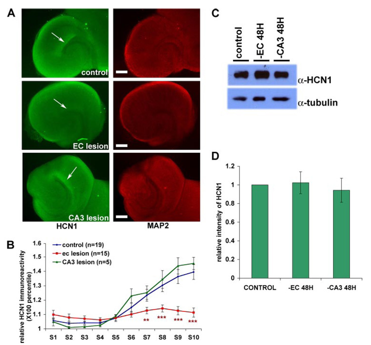
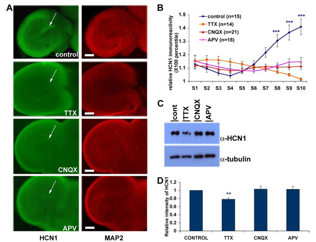
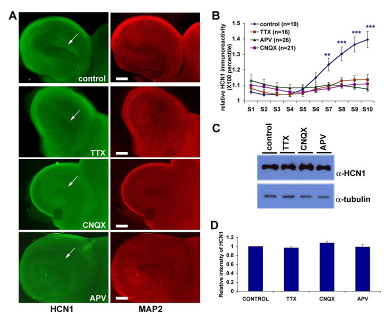
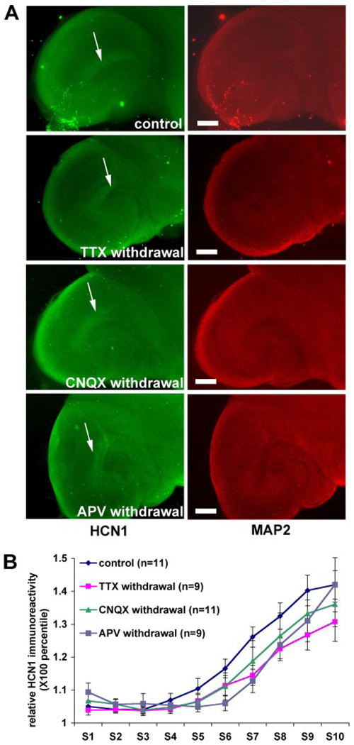
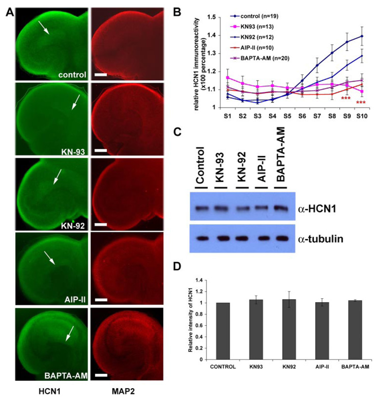
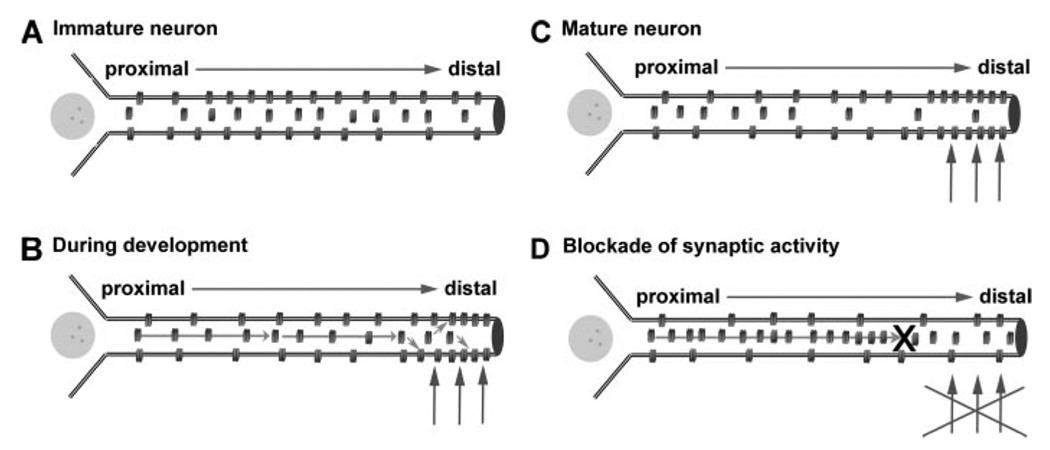
The hyperpolarization-activated cation current, I(h), plays an important role in regulating intrinsic neuronal excitability in the brain. In hippocampal pyramidal neurons, I(h) is mediated by h channels comprised primarily of the hyperpolarization-activated cyclic nucleotide-gated (HCN) channel subunits, HCN1 and HCN2. Pyramidal neuron h channels within hippocampal area CA1 are remarkably enriched in distal apical dendrites, and this unique distribution pattern is critical for regulating dendritic excitability. We utilized biochemical and immunohistochemical approaches in organotypic slice cultures to explore factors that control h channel localization in dendrites. We found that distal dendritic enrichment of HCN1 is first detectable at postnatal day 13, reaching maximal enrichment by the 3rd postnatal week. Interestingly we found that an intact entorhinal cortex, which projects to distal dendrites of CA1 but not area CA3, is critical for the establishment and maintenance of distal dendritic enrichment of HCN1. Moreover blockade of excitatory neurotransmission using tetrodotoxin, 6-cyano-7-nitroquinoxaline-2,3-dione, or 2-aminophosphonovalerate redistributed HCN1 evenly throughout the dendrite without significant changes in protein expression levels. Inhibition of calcium/calmodulin-dependent protein kinase II activity, but not p38 MAPK, also redistributed HCN1 in CA1 pyramidal neurons. We conclude that activation of ionotropic glutamate receptors by excitatory temporoammonic pathway projections from the entorhinal cortex establishes and maintains the distribution pattern of HCN1 in CA1 pyramidal neuron dendrites by activating calcium/calmodulin-dependent protein kinase II-mediated downstream signals.


Figures

FIGURE 1
FIGURE 1. gp α-HCN1 antibody is specific and sensitive in biochemical and immunohistochemical assays
A, protein extracts from COS-7 cells transfected with HCN1-expressing plasmid and mouse brains were separated by SDS-PAGE and blotted with gp α-HCN1 antibody. Our custom antibody detected a single band of ~110 kDa in transfected COS-7 cells and wild type (WT) mouse brain. No band was detected in brain extract prepared from the HCN1 knock-out mouse (Hcn1tm2Kndl). B, parasagittal sections of rat brain were immunolabeled with gp α-HCN1 antibody or antigen-preabsorbed gp α-HCN1 antibody. Antigen preabsorption eliminated immunoreactivity observed with α-HCN1, confirming specificity. C, parasagittal sections of wild type or HCN1 knock-out mouse brain were immunolabeled with gp α-HCN1 antibody. No immunoreactivity was found in HCN1 knock-out mouse brain, confirming specificity of gp α-HCN1 antibody. Scale bars, 200 µm. SO, stratum oriens; SP, stratum pyramidale; SR, stratum radiatum.
FIGURE 2
FIGURE 2. HCN1 subunits are enriched in distal dendrites of CA1 pyramidal neurons in vivo and in vitro
A, distribution of HCN1 in hippocampus of fixed rat brain. 50-µm-thick horizontal sections of adult rat brain were immunolabeled with gp α-HCN1 antibody and visualized with diaminobenzidine (left panel) or Cy3-conjugated secondary antibody (right panel). HCN1 is localized most densely in distal dendrites within SLM (arrows). B, distribution of HCN1 in dissociated hippocampal neuron culture. Dissociated neuron cultures were fixed at DIV28 and immunolabeled with α-HCN1 (left panel) and α-MAP2 (right panel) to evaluate distribution in dendrites. In contrast to brain, HCN1 is evenly distributed in the soma and along dendritic shafts in dissociated neurons (arrowheads). C, HCN1 is enriched in the distal dendrites of area CA1 in organotypic slice cultures. Organotypic slices comprising hippocampus and attached entorhinal cortex were cultured and maintained in vitro for 14 days. Immunolabeling using gp α-HCN1 (left panel) and α-MAP2 (right panel) showed enriched HCN1 subunits in distal dendritic fields of CA1 pyramidal neurons (white arrow). For further quantitation, a straight line was drawn from the cell body region to distal dendrites in CA1, and the line was divided into equal 10 sections. The section closest to the cell body layer was designated as S1, and the furthest one was designated as S10. Scale bars, 200 µm (A and C) and 20 µm (B).
FIGURE 3
FIGURE 3. Developmental changes in distribution and expression of HCN1 in organotypic slice cultures
A, organotypic slices comprising hippocampus and attached entorhinal cortex cultured from DIV1 to DIV21 were immunolabeled with α-HCN1 (green, left panels) and α-MAP2 (red, right panels). HCN1 enrichment in distal dendritic fields of CA1 pyramidal neurons becomes apparent as early as DIV6 (arrows). B, quantification of HCN1 staining in dendritic fields of CA1 pyramidal neurons. The dendritic field from soma to the SLM layer was divided into 10 segments, and the intensities of immunoreactivity of HCN1 subunits was averaged in each segment. The relative intensity of HCN1 staining in each segment was calculated by normalizing to the staining of the lowest intensity segment. At DIV1 and DIV3, HCN1 is localized in the perisomatic region, and no enrichment in distal dendrites was observed. By DIV6, distal dendritic enrichment of HCN1 is present with no significant change in distribution from DIV6 to DIV21 (DIV1, n = 9; DIV3, n = 13; DIV6, n = 9; DIV10, n = 11; DIV14, n = 15; DIV21, n = 11; **, p < 0.05; ***, p < 0.001). C, HCN1 protein expression increases during development in slice culture. Protein expression levels were analyzed by Western blotting of cultured hippocampal area CA1 extracts from DIV1 to DIV21 using gp α-HCN1 antibody. Tubulin immunoreactivity was evaluated as control for protein loading. D, quantification of HCN1 protein expression levels during development reveals that expression of HCN1 increased 3-fold from DIV1 to DIV6 and 5-fold from DIV1 to DIV14 (n = 3). Error bars represent ±S.E. Scale bars, 200 µm.
FIGURE 4
FIGURE 4. TA inputs from EC to CA1 are necessary for the establishment of HCN1 distal dendritic enrichment
A, organotypic slices were prepared at P7, and EC or CA3 was removed mechanically at the time of culturing to eliminate TA or Schaffer collateral inputs to CA1, respectively. Slices were maintained in vitro for 14 days and then immunolabeled with α-HCN1 (green, left panel) and α-MAP2 (red, right panel). In slices with both EC and CA3 attached (control) or lacking only CA3 (−CA3), HCN1 is enriched in distal dendritic fields within area CA1. In the slices without EC (−EC), HCN1 is evenly distributed throughout the dendritic field. B, quantitation of HCN1 immunoreactivity in CA1 segments of control slices as well as those lacking EC or CA3 shows distal dendritic HCN1 enrichment requires TA inputs from EC to CA1 (DIV14 control, n = 15; −EC, n = 19; −CA3, n = 13; **, p < 0.05; ***, p < 0.001). C, Western blot of CA1 extracts from DIV14 control and EC- or CA3-lesioned organotypic slice cultures probed with α-HCN1 and α-tubulin. D, intensity of the HCN1 band from Western blotting was quantitated and normalized to tubulin intensity and revealed no significant differences in area CA1 HCN1 expression levels regardless of the absence of CA3 or EC (n = 4). Arrows indicate distal dendritic field of CA1 hippocampus. Error bars represent ±S.E. Scale bars, 200 µm.
FIGURE 5
FIGURE 5. Maintenance of HCN1 distal dendritic enrichment requires the TA pathway
A, organotypic slice cultures were prepared at P7 and maintained in vitro for 14 days. At DIV14, EC or CA3 was removed mechanically, and then slices were maintained for an additional 48 h. Slices were next immunolabeled with α-HCN1 (green, left panel) and α-MAP2 (red, right panel). Removal of EC, but not CA3, resulted in loss of HCN1 distal dendritic enrichment. B, quantitation of HCN1 immunoreactivity in area CA1 dendritic fields of control, EC-, or CA3-lesioned tissue confirmed a requirement for EC to maintain HCN1 distal dendritic enrichment (DIV16 control, n = 19; EC lesion, n = 15; CA3 lesion, n = 5; **, p < 0.05; ***, p < 0.001). C, Western blot of CA1 extracts from DIV16 control and EC- or CA3-lesioned organotypic slice cultures probed with α-HCN1 and α-tubulin. D, intensity of the HCN1 bands from Western blotting was quantitated and normalized to tubulin intensity and revealed no significant changes of protein expression level within 48 h of EC or CA3 removal (control and EC lesion, n = 6; CA3 lesion, n = 4). Arrows indicate distal dendritic field of CA1 hippocampus. Error bars represent ±S.E. Scale bars, 200 µm.
FIGURE 6
FIGURE 6. Activation of ionotropic glutamate receptors is required for establishment of distal dendritic enrichment of HCN1
A, organotypic slice cultures were maintained until DIV3 and treated with the Na+ channel blocker TTX (1 µM), AMPA receptor blocker CNQX (10 µM), or NMDA receptor blocker APV (100 µM) for 11 days until DIV14. Slices were immunolabeled with gp α-HCN1 (green, left panel) and α-MAP2 (red, right panel). Distal dendritic enrichment of HCN1 was absent in area CA1 of TTX-, CNQX-, or APV-treated tissues. Note that massive cell loss was detected in EC in TTX-treated tissues but not in CNQX- or APV-treated tissues. Age-matched slices with no treatment were used as control. B, quantitation of HCN1 immunoreactivity in area CA1 dendritic fields confirmed loss of HCN1 staining in drug-treated tissues (DIV14 control, n = 15; TTX-treated, n = 14; CNQX-treated, n = 21; APV-treated, n = 18; ***, p < 0.001). C, Western blot of CA1 extracts from control (cont) or drug-treated slices were probed with α-HCN1 and α-tubulin. D, intensity of HCN1 band from Western blotting was quantitated and normalized with tubulin and revealed that expression of HCN1 was 20% decreased in TTX-treated samples but unchanged in CNQX- or APV-treated samples (n = 4; **, p < 0.05). Arrows indicate distal dendritic field of CA1 hippocampus. Error bars represent ±S.E. Scale bars, 200 µm.
FIGURE 7
FIGURE 7. Maintenance of HCN1 distal dendritic enrichment requires activation of ionotropic glutamate receptors
A, organotypic slice cultures were maintained until DIV14 and then treated with TTX (1 µM), CNQX (10 µM), or APV (100 µM) for 48 h. Slices were immunolabeled with α-HCN1 (green, left panel) and α-MAP2 (red, right panel). HCN1 staining was lost from area CA1 distal dendritic fields of slices treated with TTX, CNQX, or APV. Age-matched slices (DIV16) with no treatment were used as control. B, quantitation of HCN1 immunoreactivity in CA1 dendritic fields confirmed loss of HCN1 staining in drug-treated slices (DIV16 control, n = 19; TTX-treated, n = 16; CNQX-treated, n = 21; APV-treated, n = 26; **, p < 0.05; ***, p < 0.001). C, Western blot of CA1 extracts from control or drug-treated slices were probed with α-HCN1 and α-tubulin. D, intensity of HCN1 band from Western blotting was quantitated and normalized with tubulin and revealed that expression of HCN1 was unchanged in TTX-, CNQX-, or APV-treated samples compared with control (n = 5). Arrows indicate distal dendritic field of CA1 hippocampus. Error bars represent ±S.E. Scale bars, 200 µm.
FIGURE 8
FIGURE 8. Inhibition of HCN1 distal dendritic enrichment by Na+ channel or glutamate receptor blockade is reversible
A, slice cultures were maintained until DIV14 and then treated with TTX (1 µM), CNQX (10 µM), or APV (100 µM). After 48 h of drug treatment, drugs were washed out by medium replacement for 5 consecutive days. Slices were immunolabeled with α-HCN1 (green, left panel) and α-MAP2 (red, right panel). After washout, the HCN1 localization was restored to the same distribution as that in untreated control slices (DIV21). B, quantitation of HCN1 immunoreactivity confirmed restoration of HCN1 staining in distal CA1 dendritic fields following drug washout (DIV21 control, n = 11; TTX withdrawal, n = 9; CNQX withdrawal, n = 11; APV withdrawal, n = 9). Arrows indicate distal dendritic field of CA1 hippocampus. Error bars represent ±S.E. Scale bars, 200 µm.
FIGURE 9
FIGURE 9. Blockade of CaMKII activity redistributed HCN1 in area CA1 dendritic fields without affecting protein expression
A, organotypic slice cultures were maintained until DIV14 and then treated with the CaMKII blocker KN93 (10 µM); an inactive analogue of this molecule, KN92 (10 µM); cell-permeable CaMKII inhibitory peptide AIP-II (30 µM); or cell-permeable calcium chelator BAPTA-AM (10 µM) for 48 h. Slices were then immunolabeled with α-HCN1 (green, left panel) and α-MAP2 (red, right panel). HCN1 staining was lost from area CA1 distal dendritic fields of slices treated with KN93, AIP-II, or BAPTA-AM, but distribution was not significantly different upon treatment with KN92. Age-matched slices (DIV16) with no treatment were used as control. B, quantitation of HCN1 immunoreactivity in CA1 dendritic fields confirmed loss of HCN1 staining in KN-93-, AIP-II-, or BAPTA-AM-treated slices (DIV16 control, n = 19; KN93-treated, n = 13; KN92-treated, n = 12; AIP-II-treated, n = 10; BAPTA-AM-treated, n = 20; ***, p < 0.001). C, Western blot of CA1 extracts from control or drug-treated slices were probed with α-HCN1 and α-tubulin. D, intensity of HCN1 band from Western blotting was quantitated and normalized with tubulin and revealed that expression of HCN1 was unchanged in drug-treated slices compared with control (n = 5 for KN93 and KN92 and n = 4 for AIP-II and BAPTA-AM). Arrows indicate distal dendritic field of CA1 hippocampus. Error bars represent ±S.E. Scale bars, 200 µm.
FIGURE 10
FIGURE 10. Schematic model of activity-dependent control of HCN1 localization in the dendrites of CA1 pyramidal neurons
A, before commencement of synaptic activity, HCN1 protein is evenly distributed throughout apical dendrites. B, during development, synaptic activity (vertical arrows) through the direct EC inputs promotes trafficking of HCN1 subunits from proximal intracellular pools to the surface of distal dendrites (horizontal arrows). C, in mature neurons, h channels are enriched on the surface membrane of distal apical dendrites, and a significant proportion of HCN1 protein in proximal dendrites is intracellular, a distribution that is maintained by direct inputs from the EC. D, blockade of synaptic activity inhibits trafficking to distal dendrites (horizontal arrows), resulting in accumulation of intracellular HCN1 proximally and loss of the HCN1 gradient.

Similar articles

Cited by

References

    1. Lai HC, Jan LY. Nat. Rev. Neurosci. 2006;7:548–562. - PubMed
    1. Ludwig A, Zong X, Jeglitsch M, Hofmann F, Biel M. Nature. 1998;393:587–591. - PubMed
    1. Santoro B, Liu DT, Yao H, Bartsch D, Kandel ER, Siegelbaum SA, Tibbs GR. Cell. 1998;93:717–729. - PubMed
    1. Lorincz A, Notomi T, Tamas G, Shigemoto R, Nusser Z. Nat. Neurosci. 2002;5:1185–1193. - PubMed
    1. Notomi T, Shigemoto R. J. Comp. Neurol. 2004;471:241–276. - PubMed

Publication types

MeSH terms

Substances