. 2008 Jan 1;111(1):402-10.
doi: 10.1182/blood-2007-04-081703. Epub 2007 Sep 11.

Erythrocyte glutamine depletion, altered redox environment, and pulmonary hypertension in sickle cell disease

Affiliations

Erythrocyte glutamine depletion, altered redox environment, and pulmonary hypertension in sickle cell disease

Claudia R Morris et al. Blood. .

Abstract

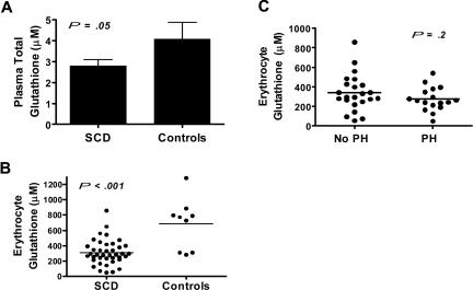
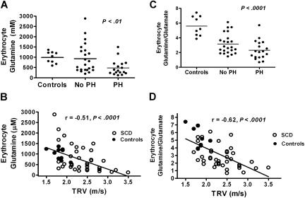
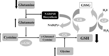
Erythrocyte glutathione depletion has been linked to hemolysis and oxidative stress. Glutamine plays an additional antioxidant role through preservation of intracellular nicotinamide adenine dinucleotide phosphate (NADPH) levels, required for glutathione recycling. Decreased nitric oxide (NO) bioavailability, which occurs in the setting of increased hemolysis and oxidative stress, contributes to the pathogenesis of pulmonary hypertension (PH) in sickle cell disease (SCD). We hypothesized that altered glutathione and glutamine metabolism play a role in this process. Total glutathione (and its precursors) and glutamine were assayed in plasma and erythrocytes of 40 SCD patients and 9 healthy volunteers. Erythrocyte total glutathione and glutamine levels were significantly lower in SCD patients than in healthy volunteers. Glutamine depletion was independently associated with PH, defined as a tricuspid regurgitant jet velocity (TRV) of at least 2.5 m/s. The ratio of erythrocyte glutamine:glutamate correlated inversely to TRV (r = -0.62, P < .001), plasma arginase concentration (r = -0.45, P = .002), and plasma-free hemoglobin level (r = -0.41, P = .01), linking erythrocyte glutamine depletion to dysregulation of the arginine-NO pathway and increased hemolytic rate. Decreased erythrocyte glutathione and glutamine levels contribute to alterations in the erythrocyte redox environment, which may compromise erythrocyte integrity, contribute to hemolysis, and play a role in the pathogenesis of PH of SCD.


Figures

Figure 1
Figure 1
Total glutathione level in patients with sickle cell disease compared with healthy controls and its association with pulmonary hypertension. Total glutathione (GSH + GSSG) levels in (A) plasma and (B) within the erythrocyte were significantly lower in patients with sickle cell disease (SCD, n = 40) compared with healthy volunteers (controls, n = 9). (C) Severity of erythrocyte glutathione deficiency was similar in patients with pulmonary hypertension (PH) when compared with those without PH (No PH)
Figure 2
Figure 2
Erythrocyte glutamine depletion in sickle cell disease and its association with pulmonary hypertension. (A) Erythrocyte glutamine levels in healthy volunteers were similar to SCD patients without pulmonary hypertension (No PH, n = 23). However, glutamine levels were significantly decreased in SCD patients with a tricuspid regurgitant jet velocity (TRV) of at least 2.5 m/s on Doppler echocardiography (PH, n = 17) compared with SCD patients without PH (P < .001) and (B) erythrocyte glutamine levels inversely correlated with TRV on Doppler echocardiography (r = -0.51, P < .0001). (C) A significant reduction in the erythrocyte glutamine:glutamate ratio is identified in SCD compared with controls, with the lowest ratios identified in patients with PH. (D) The glutamine:glutamate ratio inversely correlated with TRV on Doppler echocardiography (r = −0.62, P < .001, Figure 2D), implicating glutamine bioavailability as a novel factor in the pathophysiology of PH.
Figure 3
Figure 3
Alterations in erythrocyte glutathione (GSH) metabolism in SCD. Normal GSH metabolism: GSH is a potent antioxidant responsible for maintaining cellular redox balance. GSH is synthesized from the amino acids glutamate, cysteine (the rate-limiting substrate), and glycine. Metabolism is catalyzed sequentially by 2 cytosolic enzymes, γ-glutamylcysteine synthetase (γ-GSC) and glutathione synthetase (GS). This pathway occurs in virtually all cells, including erythrocytes, although hepatocytes are the major producer and exporter of GSH. Glutamate and cysteine are catalyzed by γ-GSC to γ-glutamyl-cysteine, which is metabolized to GSH through the actions of GS. GSH is directly oxidized to GSSG during scavenging of free radicals. It is also enzymatically oxidized by glutathione peroxidase (GPx) during the reduction of hydrogen peroxide and other peroxides. GSSG is then reduced and recycled back to GSH by the NADPH-dependent glutathione reductase (GR). During NADH and NADPH biosynthesis, glutamine is converted to glutamate. Although this is a process independent of GSH synthesis, the byproduct of this reaction is used as a substrate for GSH synthesis, while providing the essential co-enzyme for the recycling of GSH from GSSG by NADPH-dependent glutathione reductase.,, Alterations in GSH metabolism in SCD: Arrows indicate increased substrate and decreased GSH concentrations within the erythrocyte of patients with SCD. Despite more than adequate substrate, including cysteine, the rate-limiting amino acid for GSH production, GSH levels are very low in sickle erythrocytes. Further aberrations in GSH metabolism occur in patients with PH. Most notable is the erythrocyte glutamine depletion that occurs in patients with PH, compared with healthy volunteers and SCD patients without PH, while glutamate levels (significantly higher in SCD patients without PH compared with controls) begin to trend down in SCD patients with PH. Erythrocyte glutamine concentration and the glutamine:glutamate ratio inversely correlate with Doppler echocardiography tricuspid regurgitant jet velocity, a clinical marker of early mortality in SCD.

Similar articles

Cited by

References

    1. Griffith OW. Biologic and pharmacologic regulation of mammalian glutathione synthesis. Free Radic Biol Med. 1999;27:922–935. - PubMed
    1. Griffith OW. Glutathione turnover in human erythrocytes: inhibition by buthionine sulfoximine and incorporation of glycine by exchange. J Biol Chem. 1981;256:4900–4904. - PubMed
    1. Lunn G, Dale GL, Beutler E. Transport accounts for glutathione turnover in human erythrocytes. Blood. 1979;54:238–244. - PubMed
    1. Lyons J, Rauh-Pfeiffer A, Yu YM, et al. Blood glutathione synthesis rates in healthy adults receiving a sulfur amino acid-free diet. Proc Natl Acad Sci U S A. 2000;97:5071–5076. - PMC - PubMed
    1. Reid M, Badaloo A, Forrester T, et al. In vivo rates of erythrocyte glutathione synthesis in children with severe protein-energy malnutrition. Am J Physiol Endocrinol Metab. 2000;278:E405–E412. - PubMed

Publication types