. 2008 Feb 15;103(3):994-1004.
doi: 10.1002/jcb.21472.

Expression of serum amyloid A transcripts in human bone tissues, differentiated osteoblast-like stem cells and human osteosarcoma cell lines

Affiliations

Expression of serum amyloid A transcripts in human bone tissues, differentiated osteoblast-like stem cells and human osteosarcoma cell lines

Alenka Kovacevic et al. J Cell Biochem. .

Abstract

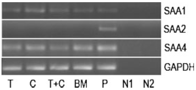
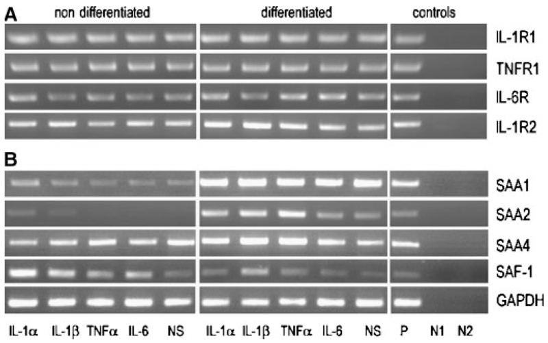
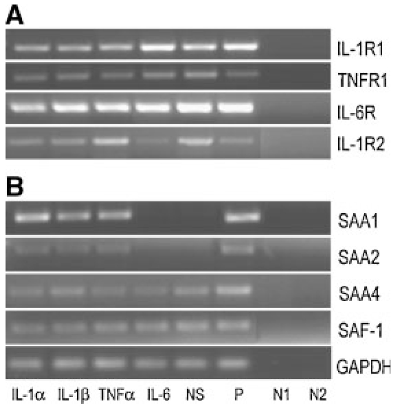
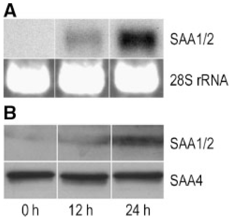
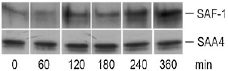
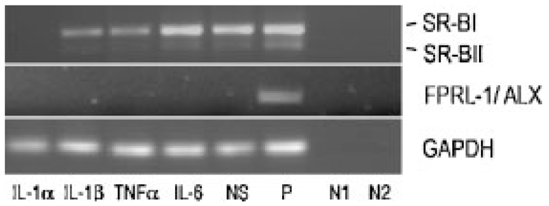
Although the liver is the primary site of cytokine-mediated expression of acute-phase serum amyloid A (SAA) protein, extrahepatic production has also been reported. Besides its role in amyloidosis and lipid homeostasis during the acute-phase, SAA has recently been assumed to contribute to bone and cartilage destruction. However, expression of SAA in human osteogenic tissue has not been studied. Therefore, we first show that SAA1 (coding for the major SAA isoform) but not SAA2 transcripts are expressed in human trabecular and cortical bone fractions and bone marrow. Next, we show expression of (i) IL-1, IL-6, and TNF receptor transcripts; (ii) the human homolog of SAA-activating factor-1 (SAF-1, a transcription factor involved in cytokine-mediated induction of SAA genes); and (iii) SAA1/2 transcripts in non-differentiated and, to a higher extent, in osteoblast-like differentiated human mesenchymal stem cells. Third, we provide evidence that human osteoblast-like cells of tumor origin (MG-63 and SAOS-2) express SAF-1 under basal conditions. SAA1/2 transcripts are expressed under basal conditions (SAOS-2) and cytokine-mediated conditions (MG-63 and SAOS-2). RT-PCR, Western blot analysis, and immunofluorescence technique confirmed cytokine-mediated expression of SAA on RNA and protein level in osteosarcoma cell lines while SAA4, a protein of unknown function, is constitutively expressed in all osteogenic tissues investigated.


Figures

Fig. 1
Fig. 1
RT-PCR of SAA transcripts in human bone: RNA was isolated from different bone preparations, reverse transcribed, and cDNAs of SAA1, SAA2, and SAA4 amplified using specific forward and reverse oligonucleotide primers (Supplement, Table I). The PCR products were separated on 1.5% agarose gels. T, trabecular bone; C, cortical bone; BM, bone marrow; P, positive control, HUH-7 cells; N1, negative control—RNA template: negative controls were done for all samples; N2, negative control—water template. To ensure that equal amount of cDNA was used as a template RT-PCR for GAPDH was made as a control. One representative experiment out of three is shown.
Fig. 2
Fig. 2
RT-PCR of cytokine receptors, SAA and SAF-1 transcripts in non-differentiated and differentiated hMSCs: hMSCs were kept in either expansion or osteogenic medium and stimulated with different cytokines (10 ng/ml) for 24 h. A: Human cytokine receptor transcripts as well as (B) SAA and SAF transcripts were amplified using specific oligonucleotide primers (Supplement, Table I). The PCR products were separated on 1.5% agarose gels. NS, non-stimulated; P, positive control: RNA was isolated from HUH-7 cells for SAF-1, SAA, cytokine receptors except IL-1R2, where RNA from THP-1 cells was used; N1, negative control— RNA template: negative controls were done for all samples; N2, negative control—water template. To ensure that equal amount of cDNA was used as a template RT-PCR for GAPDH was made as a control. One representative experiment out of three is shown.
Fig. 3
Fig. 3
RT-PCR of cytokine receptors, SAA and SAF-1 in human osteosarcoma cells: MG-63 cells were stimulated with different cytokines (10 ng/ml) for 24 h. A: Cytokine receptor transcripts as well as (B) SAA and SAF-1 transcripts were amplified using specific oligonucleotide primers (Supplement, Table I). The PCR products were separated on 1.5% agarose gels. NS, non-stimulated; P, positive control: RNA was isolated from HUH-7 cells for SAF-1, SAA, cytokine receptors except IL-1R2, where RNA from THP-1 cells was used; N1, negative control—RNA template: negative controls were done for all samples; N2, negative control—water template. To ensure that equal amount of cDNA was used as a template RT-PCR for GAPDH was made as a control. One representative experiment out of three is shown.
Fig. 4
Fig. 4
Time-dependent expression of SAA in osteosarcoma cells: MG-63 cells were stimulated with 10 ng/ml IL-1β for 12 and 24 h. A: RNA was isolated and Northern blot experiments were performed using SAA1/2 cDNA as a probe. The 28S rRNA was used as gel loading control. B: Cells were lysed and total cellular protein was subjected to SDS–PAGE and transferred to membranes. Western blot experiments were performed using sequence-specific anti-human SAA1/2 or SAA4 peptide antisera (see Materials and Methods section) as primary antibodies. One representative experiment out of three is shown.
Fig. 5
Fig. 5
Western blot of SAF-1 and SAA4 in osteosarcoma cells: MG-63 cells were stimulated with 10 ng/ml IL-1β up to 6 h in six-well plates. The cells were lysed and the nuclear fraction was isolated as described in Materials and Methods. The protein was subjected to SDS–PAGE and transferred to membranes. Western blot experiments were performed using polyclonal anti-SAF-1 antiserum or sequence-specific anti-human SAA4 peptide antisera as primary antibodies. One representative experiment out of two is shown.
Fig. 6
Fig. 6
RT-PCR of SR-BI/II and FPRL-1/ALX in human MG63 cells: Cells were stimulated with different cytokines (10 ng/ml) for 24 h. Human SR-BI/II and FPRL-1/ALX transcripts were amplified using specific oligonucleotide primers (Supplement, Table I). The PCR products were separated on 1% agarose gels. NS, non-stimulated; P, positive control: RNA was isolated from differentiated THP-1 cells for SR-BI/II, for FPRL-1/ALX, HUH-7 genomic DNA was used; N1, negative control—RNA template: negative controls were done for all samples; N2, negative control—water template. To ensure that equal amount of cDNA was used as a template RT-PCR for GAPDH was made as a control. One representative experiment out of three is shown.

References

    1. Artl A, Marsche G, Lestavel S, Sattler W, Malle E. Role of serum amyloid A during metabolism of acute-phase HDL by macrophages. Arterioscler Thromb Vasc Biol. 2000;20:763–772. - PubMed
    1. Badolato R, Wang JM, Murphy WJ, Lloyd AR, Michiel DF, Bausserman LL, Kelvin DJ, Oppenheim JJ. Serum amyloid A is a chemoattractant: Induction of migration, adhesion, and tissue infiltration of monocytes and polymorphonuclear leukocytes. J Exp Med. 1994;180:203–209. - PMC - PubMed
    1. Badolato R, Johnston JA, Wang JM, McVicar D, Xu LL, Oppenheim JJ, Kelvin DJ. Serum amyloid A induces calcium mobilization and chemotaxis of human monocytes by activating a pertussis toxin-sensitive signaling pathway. J Immunol. 1995;155:4004–4010. - PubMed
    1. Baranova IN, Vishnyakova TG, Bocharov AV, Kurlander R, Chen Z, Kimelman ML, Remaley AT, Csako G, Thomas F, Eggerman TL, Patterson AP. Serum amyloid A binding to CLA-1 (CD36 and LIMPII analogous-1) mediates serum amyloid A protein-induced activation of ERK1/2 and p38 mitogen-activated protein kinases. J Biol Chem. 2005;280:8031–8040. - PubMed
    1. Biran H, Friedman N, Neumann L, Pras M, Shainkin-Kestenbaum R. Serum amyloid A (SAA) variations in patients with cancer: Correlation with disease activity, stage, primary site, and prognosis. J Clin Pathol. 1986;39:794–797. - PMC - PubMed

Publication types

MeSH terms