

In vivo magnetic resonance imaging of vascularization in islet transplantation
- PMID: 17850231
- PMCID: PMC2094099
- DOI: 10.1111/j.1432-2277.2007.00550.x
In vivo magnetic resonance imaging of vascularization in islet transplantation
Abstract
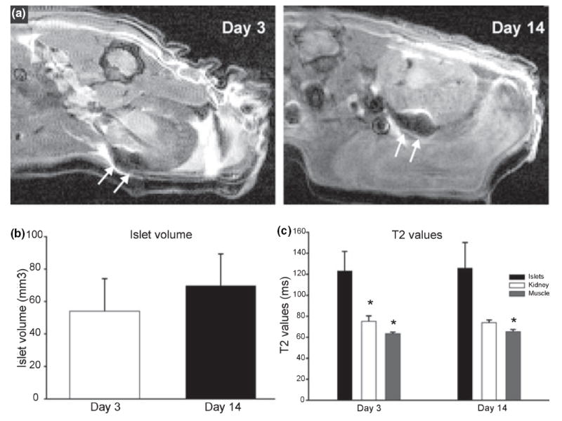
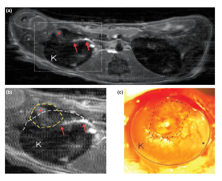
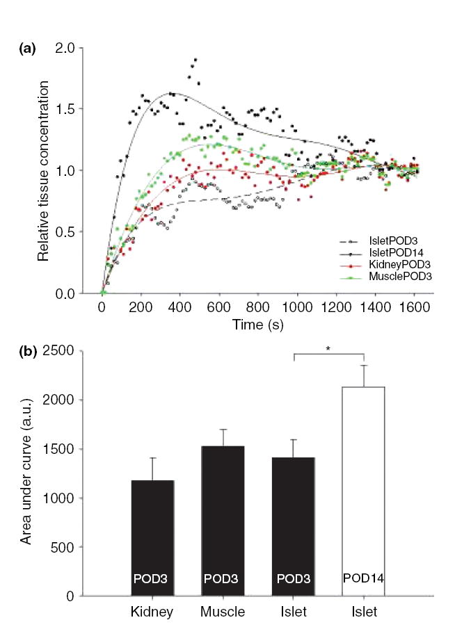
To evaluate changes in neovascularization of transplanted islets in vivo, dynamic contrast (gadolinium) enhanced magnetic resonance imaging (MRI) was used. Both iron (Feridex)-labeled and unlabeled syngeneic murine subcapsular islet grafts were studied. Differences in dynamic contrast enhancement of islet grafts were quantified after gadolinium injection at post-transplant days 3 and 14. Normalized contrast concentrations at day 14 in transplanted islets were increased relative with that on day 3. Time to peak contrast enhancement was faster by 12 min at day 14 compared to day 3 islets (while kidney and muscle peak times remained the same). Areas under the curve for contrast concentration versus time plots were larger in 14-day relative to 3-day islet grafts. In conclusion, noninvasive assessment of neovascularization is achievable. In vivo dynamic contrast-enhanced MRI can be used to detect and quantify changes in vascularization following islet transplantation. This technique may be useful in developing pro-angiogenic strategies to improve the transplantation outcome in experimental and clinical settings.
Figures
References
-
- Ryan EA, Lakey JR, Paty BW, et al. Successful islet transplantation: continued insulin reserve provides long-term glycemic control. Diabetes. 2002;51:2148. - PubMed
-
- Davalli AM, Ogawa Y, Ricordi C, Scharp DW, Bonner-Weir S, Weir GC. A selective decrease in the beta cell mass of human islets transplanted into diabetic nude mice. Transplantation. 1995;59:817. - PubMed
-
- Shapiro AM, Ryan EA, Lakey JR. Diabetes. Islet cell transplantation. Lancet. 2001;358(Suppl):S21. - PubMed
-
- Carlsson PO, Palm F, Andersson A, Liss P. Markedly decreased oxygen tension in transplanted rat pancreatic islets irrespective of the implantation site. Diabetes. 2001;50:489. - PubMed
-
- Wang W, Upshaw L, Strong DM, Robertson RP, Reems J. Increased oxygen consumption rates in response to high glucose detected by a novel oxygen biosensor system in non-human primate and human islets. J Endocrinol. 2005;185:445. - PubMed
Publication types
MeSH terms
Substances
Grants and funding
LinkOut - more resources
Full Text Sources
Other Literature Sources
Medical
