. 2007 Oct;66(2):329-40.
doi: 10.1111/j.1365-2958.2007.05908.x. Epub 2007 Sep 10.

RrgA is a pilus-associated adhesin in Streptococcus pneumoniae

Affiliations

RrgA is a pilus-associated adhesin in Streptococcus pneumoniae

A L Nelson et al. Mol Microbiol. 2007 Oct.

Abstract

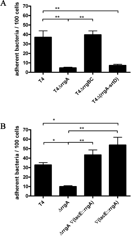
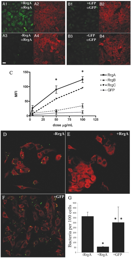
Adherence to host cells is important in microbial colonization of a mucosal surface, and Streptococcus pneumoniae adherence was significantly enhanced by expression of an extracellular pilus composed of three subunits, RrgA, RrgB and RrgC. We sought to determine which subunit(s) confers adherence. Bacteria deficient in RrgA are significantly less adherent than wild-type organisms, while overexpression of RrgA enhances adherence. Recombinant monomeric RrgA binds to respiratory cells, as does RrgC with less affinity, and pre-incubation of epithelial cells with RrgA reduces adherence of wild-type piliated pneumococci. Non-adherent RrgA-negative, RrgB- and RrgC-positive organisms produce pili, suggesting that pilus-mediated adherence is due to expression of RrgA, rather than the pilus backbone itself. In contrast, RrgA-positive strains with disrupted rrgB and rrgC genes exhibit wild-type adherence despite failure to produce pili by Western blot or immunoelectron microscopy. The density of bacteria colonizing the upper respiratory tract of mice inoculated with piliated RrgA-negative pneumococci was significantly less compared with wild-type; in contrast, non-piliated pneumococci expressing non-polymeric RrgA had similar numbers of bacteria in the nasopharynx as piliated wild-type bacteria. These data suggest that RrgA is central in pilus-mediated adherence and disease, even in the absence of polymeric pilus production.


Figures

Fig. 1
Fig. 1
Expression of rrgA confers pilus-mediated adherence to human respiratory epithelial cells. A. rrgA, but not rrgB and rrgC, is necessary for pilus-mediated adherence to epithelial cells. Adherence of wild-type piliated TIGR4 (‘T4’) to A549 human respiratory epithelial cells was significantly greater than both non-piliated T4Δ(rrgA-srtD) compared with T4 deficient in the rrgA gene (‘T4ΔrrgA’). Adherence of T4 deficient in both rrgB and rrgC (‘T4ΔrrgBC’) was not notably different from wild-type organisms, but was significantly greater compared with T4ΔrrgA. Repeated-measure anova of data collected from three independent determinations indicates statistically significant differences within experimental conditions. Post hoc Bonferroni analyses identify specific significant differences: **P< 0.01. B. rrgA expression determines adherence to epithelial cells. T4 deficient in rrgA are significantly deficient in adherence (‘ΔrrgA’), compared with wild-type (T4), while trans-complementation restores wild-type adherence [‘ΔrrgA∇(lacE::rrgA’)]. Introduction of a second copy of rrgA inserted in trans in the lacE locus [‘∇(lacE::rrgA’] results in significant enhancement of adherence over wild-type levels. Statistical analyses were performed with repeated-measure anova of data collected from three independent determinations. Post hoc Bonferroni analyses identify specific significant differences: *P< 0.01 and **P< 0.001.
Fig. 2
Fig. 2
Recombinant RrgA and RrgC proteins bind to human respiratory epithelial cells. A–C. Recombinant RrgA binds directly to A549 respiratory epithelial cells. Cells were incubated with 100 μg ml−1 of purified RrgA (A1 and A2, ‘+RrgA’) or GFP (B1 and B2, ‘+GFP’) in DMEM culture medium, or medium alone (A3–4, ‘-RrgA’; B3–4, ‘-GFP’) for 2 h at 4°C. Cells were fixed and stained with anti-RrgA and anti-GFP antibodies (A1–4, ‘αRrgA’; B1–4, ‘αGFP’) and phalloidin, which served as a control to demonstrate the presence of cells (A2, A4, B2 and B4). Imaging was performed with a confocal microscope. Scale bar is 20 μm. C. RrgA and RrgC bind to A549 cells in a dose-dependent manner. A549 cells were mixed in suspension with either medium alone (0 μg ml−1) or three concentrations (5, 50 and 100 μg ml−1) of pilus subunits RrgA (squares with solid black lines), RrgB (triangles with dashed grey lines), RrgC (upside-down triangles with dashed black lines), or GFP protein (diamond with solid grey lines), incubated for 2 h at 4°C, stained with antisera specific to each protein, and detected with Alexa Fluor 488-conjugated secondaries. Cells were analysed with a FACS-Calibur flow cytometer, and the net mean fluorescence intensity for each population was calculated from three independent experiments. Significant differences were detected by repeated-measure anova (P< 0.0001), and both RrgA and RrgC binding was significantly different from RrgB and GFP binding at 50 and 100 μg ml−1 by post hoc Bonferroni analysis (*P < 0.001). D–G. Pre-incubation of A549 cells with purified RrgA protein inhibits pilus-mediated adherence. A549 cells were pre-incubated with media alone (D), media containing 100 μg ml−1 of RrgA (E), or 100 μg ml−1 of GFP (F). After pre-incubation, A549 monolayers were infected with S. pneumoniae strain T4. Cells were stained with phalloidin (red) anti-S. pneumoniae capsule antibody (green), and imaged with a confocal microscope. RrgA pre-incubation inhibits the adherence of strain T4 to the A549 cells (E versus D), while the negative control GFP protein does not (F versus D). Scale bar is 20 μm. Adherent bacteria were counted, and the number of bacteria adherent to 100 A549 cells is shown in G (n = 6 fields, *P= 0.0002, **P= 0.8).
Fig. 3
Fig. 3
The pneumococcal pilus is expressed in the absence of RrgA. Production of polymeric high-molecular-weight extracellular pili in isogenic sets of pneumococci was evaluated by preparation of cell wall-associated proteins and immunoblotting for pilus subunits RrgA (A), RrgB (B) and RrgC (C). In all cases, cell wall proteins were separated by 4–12% gradient SDS-PAGE, electrotransferred to a PVDF membrane, and probed for pilus subunits. Approximate molecular weight in kDa, based on marker proteins, are indicated on left. A. Immunoblotting for RrgA in wild-type T4, T4ΔrrgA (‘ΔrrgA’), T4ΔrrgBC (‘ΔrrgBC’), and T4Δ(rrgA-srtD) (‘rrgA-srtD’). B. Immunoblotting for RrgB in wild-type piliated T4, T4ΔrrgA, T4ΔrrgBC and T4Δ(rrgA-srtD), labelled as in A. C. Immunoblotting for RrgC in wild-type piliated T4, T4ΔrrgA, T4ΔrrgBC, and T4Δ(rrgA-srtD), labelled as in A. These data demonstrate that extracellular RrgB-positive, RrgC-positive pili are formed in the absence of rrgA expression, but not in the absence of expression of rrgB and rrgC. In the absence of RrgB and RrgC, RrgA is found as a 100 kDa monomer.
Fig. 4
Fig. 4
The pneumococcal pilus is observed in the absence of RrgA expression. A. Immunogold detection of RrgA (10 nm gold particles, indicated by open arrow) and RrgB (5 nm gold particle, indicated by arrowheads) in wild-type T4. B. Immunogold detection of RrgA (10 nm gold particles, open arrow), RrgB (5 nm gold particles, arrowhead) and RrgC (15 nm gold particles, closed arrow) in T4ΔrrgA. Scale bar is 200 nm. No RrgA was detected in T4ΔrrgA preparations. Note that polymeric RrgB- and RrgC-positive pili are present in T4 strains in the presence (A) or absence (B) of rrgA expression. C. Immunogold detection of RrgA (10 nm gold particle, open arrow), RrgB (5 nm gold particles, arrowhead), and RrgC (15 nm gold particles, closed arrow), in T4ΔrrgBC. Scale bar is 200 nm. No RrgB or RrgC was detected in T4ΔrrgBC preparations. Note that RrgA is found in extracellular pili in wild-type T4, but in association with the cell wall in T4ΔrrgBC, where no pili are observed.
Fig. 5
Fig. 5
RrgA is involved in pilus-mediated colonization of the upper respiratory tract in mice. Mice were challenged intranasally with a low dose of T4, T4ΔrrgA or T4ΔrrgBC (c. 7 × 10⁴ cfu per mouse). Bacterial density in the nasopharynx was determined 7 days post infection. Mice infected with T4ΔrrgA had significantly fewer bacteria in the nasopharynx than mice infected with T4 or T4ΔrrgBC (*P< 0.01, n = 3 independent replicates of 10 mice per group per replicate, using the Kruskal–Wallis test with Dunn's post testing).

References

    1. Aaberge IS, Eng J, Lermark G, Lovik M. Virulence of Streptococcus pneumoniae in mice: a standardized method for preparation and frozen storage of the experimental bacterial inoculum. Microb Pathog. 1995;18:141–152. - PubMed
    1. Abbot EL, Smith WD, Siou GP, Chiriboga C, Smith RJ, Wilson JA, et al. Pili mediate specific adhesion of Streptococcus pyogenes to human tonsil and skin. Cell Microbiol. 2007;9:1822–1833. - PubMed
    1. Abeyta M, Hardy GG, Yother J. Genetic alteration of capsule type but not PspA type affects accessibility of surface-bound complement and surface antigens of Streptococcus pneumoniae. Infect Immun. 2003;71:218–225. - PMC - PubMed
    1. Albiger B, Sandgren A, Katsuragi H, Meyer-Hoffert U, Beiter K, Wartha F, et al. Myeloid differentiation factor 88-dependent signalling controls bacterial growth during colonization and systemic pneumococcal disease in mice. Cell Microbiol. 2005;7:1603–1615. - PubMed
    1. Barocchi MA, Ries J, Zogaj X, Hemsley C, Albiger B, Kanth A, et al. A pneumococcal pilus influences virulence and host inflammatory responses. Proc Natl Acad Sci USA. 2006;103:2857–2862. - PMC - PubMed

Publication types

MeSH terms