. 2007 Sep 12:7:84.
doi: 10.1186/1471-2180-7-84.

Salmonella induces prominent gene expression in the rat colon

Affiliations

Salmonella induces prominent gene expression in the rat colon

Wendy Rodenburg et al. BMC Microbiol. .

Abstract

Background: Salmonella enteritidis is suggested to translocate in the small intestine. In vivo it induces gene expression changes in the ileal mucosa and Peyer's patches. Stimulation of Salmonella translocation by dietary prebiotics fermented in colon suggests involvement of the colon as well. However, effects of Salmonella on colonic gene expression in vivo are largely unknown. We aimed to characterize time dependent Salmonella-induced changes of colonic mucosal gene expression in rats using whole genome microarrays. For this, rats were orally infected with Salmonella enteritidis to mimic a foodborne infection and colonic gene expression was determined at days 1, 3 and 6 post-infection (n = 8 rats per time-point). As fructo-oligosaccharides (FOS) affect colonic physiology, we analyzed colonic mucosal gene expression of FOS-fed versus cellulose-fed rats infected with Salmonella in a separate experiment. Colonic mucosal samples were isolated at day 2 post-infection.

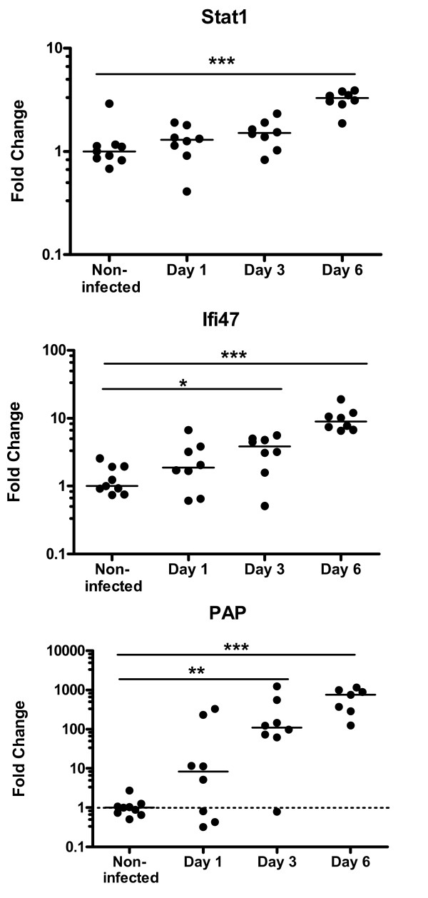
Results: Salmonella affected transport (e.g. Chloride channel calcium activated 6, H+/K+ transporting Atp-ase), antimicrobial defense (e.g. Lipopolysaccharide binding protein, Defensin 5 and phospholipase A2), inflammation (e.g. calprotectin), oxidative stress related genes (e.g. Dual oxidase 2 and Glutathione peroxidase 2) and Proteolysis (e.g. Ubiquitin D and Proteosome subunit beta type 9). Furthermore, Salmonella translocation increased serum IFN gamma and many interferon-related genes in colonic mucosa. The gene most strongly induced by Salmonella infection was Pancreatitis Associated Protein (Pap), showing >100-fold induction at day 6 after oral infection. Results were confirmed by Q-PCR in individual rats. Stimulation of Salmonella translocation by dietary FOS was accompanied by enhancement of the Salmonella-induced mucosal processes, not by induction of other processes.

Conclusion: We conclude that the colon is a target tissue for Salmonella, considering the abundant changes in mucosal gene expression.


Figures

Figure 1
Figure 1
Sum of urinary nitrate and nitrite (NOx) excretion in the non-infected (○), infected (■) groups of the time course infection study (A). And the urinary NOx excretion in the cellulose infected (■) and in the fructo-oligosaccharide (FOS) infected (▲) groups in the dietary infection study (B). Infected rats were orally challenged with S. enteritidis on day 0. Results are expressed as mean ± SEM (n = 8 in the time course infection study and n = 6 in the dietary infection study). * p < 0.05.
Figure 2
Figure 2
The number of differentially expressed genes with a fold change greater than 2 in colon mucosa of rats at days 1, 3 or 6 after oral infection with Salmonella or control treatment.
Figure 3
Figure 3
Serum IFNγ levels before and after infection (days 1, 3 and 6 p.i.). Each dot represents an individual rat. Group medians are presented by a black line. * p < 0.05, ***p < 0.001.
Figure 4
Figure 4
Individual expression of two interferon-related genes and Pap in the colon mucosa at different time points after Salmonella infection or control treatment. Genes expression is quantified by Q-PCR, using Rps26 as reference gene (using Arf1 as reference gene showed similar results; data not shown). Each dot represents an individual rat. Dotted line indicate lowest mRNA standard. Medians are presented by a black line. The median value of the uninfected group was set at 1.0. Y-axis is at log10 scale. * p < 0.05, **p < 0.01, ***p < 0.001.
Figure 5
Figure 5
Expression level of most consistent Salmonella-target genes in colon mucosa of rats fed a cellulose diet or a FOS diet. The gene expression is obtained from micro array analysis of pooled colonic mucosa samples collected at day 2 post-infection. Each dot represents a gene. The median value of each gene in the uninfected group is set to 1.0. Y-axis is at log2scale. ***p < 0.001.

Similar articles

Cited by

References

    1. Barrow PA. Salmonella infections: immune and non-immune protection with vaccines. Avian Pathol. 2007;36:1–13. doi: 10.1080/03079450601113167. - DOI - PubMed
    1. Herikstad H, Motarjemi Y, Tauxe RV. Salmonella surveillance: a global survey of public health serotyping. Epidemiol Infect. 2002;129:1–8. doi: 10.1017/S0950268802006842. - DOI - PMC - PubMed
    1. Havelaar AH, Garssen J, Takumi K, Koedam MA, Dufrenne JB, van Leusden FM, de La Fonteyne L, Bousema JT, Vos JG. A rat model for dose-response relationships of Salmonella Enteritidis infection. J Appl Microbiol. 2001;91:442–452. doi: 10.1046/j.1365-2672.2001.01399.x. - DOI - PubMed
    1. Naughton PJ, Grant G, Spencer RJ, Bardocz S, Pusztai A. A rat model of infection by Salmonella typhimurium or Salm. enteritidis. J Appl Bacteriol. 1996;81:651–656. - PubMed
    1. Rodenburg W, Bovee-Oudenhoven IMJ, Kramer E, van der Meer R, Keijer J. Gene expression response of the rat small intestine following oral Salmonella infection. Physiological Genomics. 2007;30:123–133. doi: 10.1152/physiolgenomics.00190.2006. - DOI - PubMed

Publication types

MeSH terms