Comparative Study
. 2007;9(5):R59.
doi: 10.1186/bcr1765.

Aging impacts transcriptomes but not genomes of hormone-dependent breast cancers

Affiliations
Comparative Study

Aging impacts transcriptomes but not genomes of hormone-dependent breast cancers

Christina Yau et al. Breast Cancer Res. 2007.

Abstract

Introduction: Age is one of the most important risk factors for human malignancies, including breast cancer; in addition, age at diagnosis has been shown to be an independent indicator of breast cancer prognosis. Except for inherited forms of breast cancer, however, there is little genetic or epigenetic understanding of the biological basis linking aging with sporadic breast cancer incidence and its clinical behavior.

Methods: DNA and RNA samples from matched estrogen receptor (ER)-positive sporadic breast cancers diagnosed in either younger (age <or= 45 years) or older (age >or= 70 years) Caucasian women were analyzed by array comparative genomic hybridization and by expression microarrays. Array comparative genomic hybridization data were analyzed using hierarchical clustering and supervised age cohort comparisons. Expression microarray data were analyzed using hierarchical clustering and gene set enrichment analysis; differential gene expression was also determined by conditional permutation, and an age signature was derived using prediction analysis of microarrays.

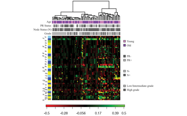
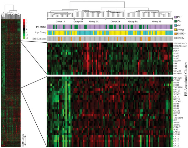
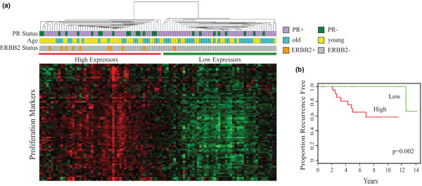
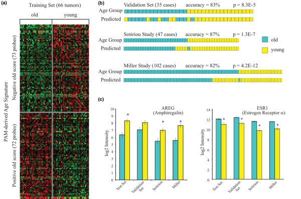
Results: Hierarchical clustering of genome-wide copy-number changes in 71 ER-positive DNA samples (27 younger women, 44 older women) demonstrated two age-independent genotypes; one with few genomic changes other than 1q gain/16q loss, and another with amplifications and low-level gains/losses. Age cohort comparisons showed no significant differences in total or site-specific genomic breaks and amplicon frequencies. Hierarchical clustering of 5.1 K genes variably expressed in 101 ER-positive RNA samples (53 younger women, 48 older women) identified six transcriptome subtypes with an apparent age bias (P < 0.05). Samples with higher expression of a poor outcome-associated proliferation signature were predominantly (65%) younger cases. Supervised analysis identified cancer-associated genes differentially expressed between the cohorts; with younger cases expressing more cell cycle genes and more than threefold higher levels of the growth factor amphiregulin (AREG), and with older cases expressing higher levels of four different homeobox (HOX) genes in addition to ER (ESR1). An age signature validated against two other independent breast cancer datasets proved to have >80% accuracy in discerning younger from older ER-positive breast cancer cases with characteristic differences in AREG and ESR1 expression.

Conclusion: These findings suggest that epigenetic transcriptome changes, more than genotypic variation, account for age-associated differences in sporadic breast cancer incidence and prognosis.


Figures

Figure 1
Figure 1
Hierarchical clustering of primary estrogen receptor-positive breast cancers based on genome-wide DNA copy number aberrations. Unsupervised hierarchical clustering of 71 primary estrogen receptor-positive breast cancers, diagnosed in younger women (age ≤ 45 years) or older women (age ≥ 70 years), based on genome-wide DNA copy number aberrations. As previously reported for BAC-based array comparative genomic hybridization analyses of human breast cancer samples [5], columns represent individual tumor samples and rows represent individual genome probes (BAC clones), ordered by chromosome and genome position with 1pter at the top and 22qter at the bottom. Chromosome p-arms and q-arms are shown as different shades of the same color (blue = odd numbered chromosomes, yellow = even numbered chromosomes). As indicated in the color scale at the bottom, genome copy number losses are indicated in red (-0.5) and copy number gains are indicated in green (0.5). Yellow dots represent high-level genomic amplifications. Colored and grey-toned upper bars identify the age cohort, progesterone receptor (PR) status, nodal status and grade status of the estrogen receptor-positive samples in each column. The dendrogram shows unsupervised classification of the 71 samples into two primary clusters and four secondary clusters, with no significant cluster bias according to age, PR status, nodal status or grade status (P > 0.3, Fisher exact test).
Figure 2
Figure 2
Hierarchical clustering of primary estrogen receptor-positive breast cancers based on genome-wide microarray profiling. Unsupervised hierarchical clustering of 101 primary estrogen receptor (ER)-positive breast cancers, diagnosed in younger women (age ≤ 45 years) or older women (age ≥ 70 years), based on genome-wide microarray profiling of 6,632 variably expressed probes (~5.1 K unique genes). The cluster dendrogram defines six different transcriptome subtypes of ER-positive breast cancers (Group 1A, 1B, 2A, 2B, 3A, 3B), with significant age biases (P < 0.05) but not biased by progesterone receptor (PR) status or ERBB2 status; horizontal colored bars identify the age cohort, PR status and ERBB2 status of the ER-positive samples in each column. The vertical red–green color scale shows log2 ratios from mean centered gene expression levels. Magnified views show ESR1-containing (ER-associated) probe sets within the entire cluster diagram.
Figure 3
Figure 3
Estrogen receptor-positive breast cancer subsets by gene set enrichment analysis. Assessment of estrogen receptor (ER)-positive breast cancer subsets by gene set enrichment analysis (GSEA) for specific gene signatures. (a) Unsupervised clustering of the 101 primary ER-positive breast cancers shown in Figure 2 based only on expression of the 71-gene proliferation signature shown to be significant by GSEA, revealing two major clusters (high expressors and low expressors of proliferation signature) with significant biases in age and ERBB2 status; horizontal colored bars identify the age cohort, progesterone receptor (PR) status and ERBB2 status of the samples in each column. (b) Kaplan–Meier plots of recurrence events among the 54 ER-positive cases with known clinical follow-up, dichotomized by high (red) or low (green) expression of the 71-gene proliferation signature, with significance determined by log-rank analysis.
Figure 4
Figure 4
Prediction analysis of microarrays-derived age signature validated against independent estrogen receptor-positive breast cancer datasets. (b) University of California San Francisco (UCSF) RNA sample set 2 (n = 66, younger women and older women) was used to train prediction analysis of microarrays (PAM) and to derive a 145-probe (128-gene) age cohort classifying signature, arranged in ascending order of the PAM score for cases in the older cohort. (b) Actual and signature-predicted age cohort designations for the validating UCSF RNA sample set 1 (n = 35) and two external validating datasets restricted to estrogen receptor-positive cases with identical age cohort characteristics: Sotiriou and colleagues [48] and Miller and colleagues [47]. Prediction accuracies are indicated, with Fisher's exact test P values presented for significance. (c) Age-signature-defined subsets from all four sample datasets show similar differences in log2 expression levels (mean ± standard deviation) of AREG and ESR1.

References

    1. Edwards B, Howe HL, Ries L, Thun M, Rosenberg H, Wingo P, Jemal A, Feigal E. Annual Report to the Nation on the Status of Cancer, 1973–1999 featuring implications of age and aging on US cancer burden. Cancer. 2000;94:2766–2792. doi: 10.1002/cncr.10593. - DOI - PubMed
    1. Smigal C, Jemal A, Ward E, Cokkinides V, Smith R, Howe HL, Thun M. Trends in breast cancer by race and ethnicity: update 2006. CA Cancer J Clin. 2006;56:168–183. - PubMed
    1. DevCan – probability of developing or dying of cancer software http://www.srab.cancer.gov/devcan
    1. Benz CC, Campisi J, Cohen HJ, Ershler WB, Haubein L, Irminger-Finger I. Meeting report: translational research at the aging and cancer interface. Cancer Res. 2007;67:4560–4563. doi: 10.1158/0008-5472.CAN-07-1236. - DOI - PubMed
    1. Geigl JB, Langer S, Barwisch S, Pfleghaar K, Lederer G, Speicher MR. Analysis of gene expression patterns and chromosomal changes associated with aging. Cancer Res. 2004;64:8550–8557. doi: 10.1158/0008-5472.CAN-04-2151. - DOI - PubMed

Publication types

MeSH terms