

Evidence of altered epidermal nerve fiber morphology in adults with self-injurious behavior and neurodevelopmental disorders
- PMID: 17850969
- PMCID: PMC3533420
- DOI: 10.1016/j.pain.2007.07.022
Evidence of altered epidermal nerve fiber morphology in adults with self-injurious behavior and neurodevelopmental disorders
Abstract
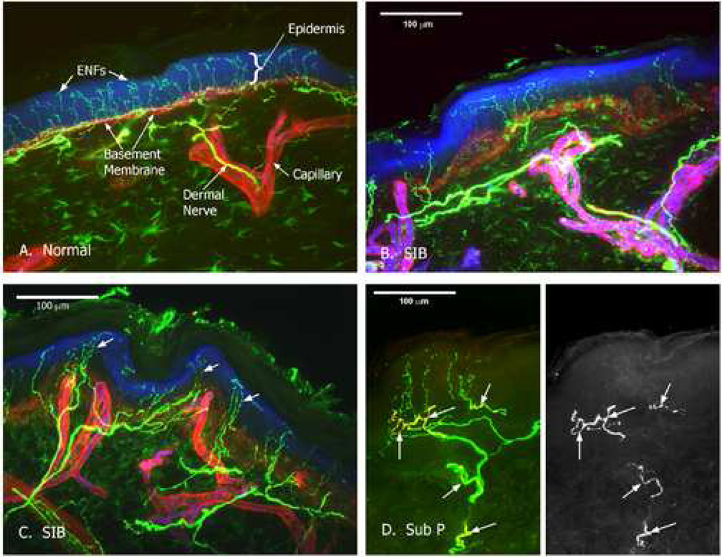
The purpose of this preliminary study was to examine the morphology and neuropeptide density of epidermal nerve fibers quantified through skin biopsy samples from three adults with neurodevelopmental disorders and chronic self-injurious behavior (SIB) secondary to mental retardation compared with non-SIB normal IQ controls. A cross-sectional design was used with 3mm punch skin biopsies collected from each participant from non-self-injurious body sites and compared with site-matched existing normal control skin samples. The study was conducted at an outpatient clinic. The primary dependent measure for the morphology analyses was the coefficient of variation (CV) to quantify the mean gap length between epidermal nerve fibers for each subject. Visual microscopic examination and quantitative analysis of the microscopy images suggested there were morphological abnormalities (increased CV) in the epidermal nerve fibers among the chronic SIB cases. Substance P (SP) fiber density was increased with 2-3 times as many fibers in SIB subjects as control subjects. Additional empirical work is needed to clarify the relation between sensory innervation of the skin and self-injury to improve assessment and treatment outcomes.
Figures
Similar articles
-
Degranulated mast cells in the skin of adults with self-injurious behavior and neurodevelopmental disorders.Brain Behav Immun. 2009 Mar;23(3):365-70. doi: 10.1016/j.bbi.2008.11.003. Epub 2008 Nov 27. Brain Behav Immun. 2009. PMID: 19084591
-
Altered diurnal pattern of salivary substance P in adults with developmental disabilities and chronic self-injury.Am J Ment Retard. 2003 Jan;108(1):13-8. doi: 10.1352/0895-8017(2003)108<0013:ADPOSS>2.0.CO;2. Am J Ment Retard. 2003. PMID: 12475363
-
Skin blister and skin biopsy to quantify epidermal nerves: a comparative study.Neurology. 2009 Apr 7;72(14):1205-10. doi: 10.1212/01.wnl.0000340984.74563.1c. Epub 2008 Dec 17. Neurology. 2009. PMID: 19092108
-
The cutaneous nerve biopsy: technical aspects, indications, and contribution.Handb Clin Neurol. 2013;115:171-88. doi: 10.1016/B978-0-444-52902-2.00010-2. Handb Clin Neurol. 2013. PMID: 23931780 Review.
-
Assessment of epidermal nerve fibers: a new diagnostic and predictive tool for peripheral neuropathies.J Neuropathol Exp Neurol. 2007 Dec;66(12):1059-73. doi: 10.1097/nen.0b013e31815c8989. J Neuropathol Exp Neurol. 2007. PMID: 18090915 Review.
Cited by
-
Neuronal cytoskeletal gene dysregulation and mechanical hypersensitivity in a rat model of Rett syndrome.Proc Natl Acad Sci U S A. 2017 Aug 15;114(33):E6952-E6961. doi: 10.1073/pnas.1618210114. Epub 2017 Jul 31. Proc Natl Acad Sci U S A. 2017. PMID: 28760966 Free PMC article.
-
Initially intact neural responses to pain in autism are diminished during sustained pain.Autism. 2018 Aug;22(6):669-683. doi: 10.1177/1362361317696043. Epub 2017 May 17. Autism. 2018. PMID: 28513186 Free PMC article.
-
Dermatomal scratching after intramedullary quisqualate injection: correlation with cutaneous denervation.J Pain. 2008 Nov;9(11):999-1005. doi: 10.1016/j.jpain.2008.05.010. Epub 2008 Jul 10. J Pain. 2008. PMID: 18619906 Free PMC article.
-
A systematic review of risk for the development and persistence of self-injurious behavior in intellectual and developmental disabilities.Clin Psychol Rev. 2022 Jun;94:102158. doi: 10.1016/j.cpr.2022.102158. Epub 2022 Apr 22. Clin Psychol Rev. 2022. PMID: 35580423 Free PMC article.
-
Delineating subtypes of self-injurious behavior maintained by automatic reinforcement.J Appl Behav Anal. 2015 Sep;48(3):523-43. doi: 10.1002/jaba.236. Epub 2015 Jul 29. J Appl Behav Anal. 2015. PMID: 26223959 Free PMC article.
References
-
- Albrecht PJ, Hines S, Eisenberg E, Pud D, Finaly DR, Connolly MK, Pare M, Davar G, Rice FL. Pathologic alterations of cutaneous innervation and vasculature in affected limbs from patients with complex regional pain syndrome. Pain. 2006;120:244–266. - PubMed
-
- Barrera FJ, Teodoro JM, Selmeci T, Madappuli A. Self-injury, pain, and the endorphin theory. J Dev Phys Disab. 1994;6:169–192.
-
- Breau L, Boll P, McKay A, Symons FJ, et al. Pain and self-injurious behavior in neurologically impaired children. J Ped. 2003;142:498–503. - PubMed
-
- Dan Y, Mooney RD. Sensory systems. Curr Op Neurobio. 2006;16:359–362.
-
- Dou YC, Hagstromer L, Emtestam L, Johannson O. Increased nerve growth factor and its receptors in atopic dermatitis: an immunohistochemical study. Arch Dermatol Res. 2006;296:31–37. - PubMed
Publication types
MeSH terms
Substances
Grants and funding
LinkOut - more resources
Full Text Sources
Medical
Miscellaneous
