

Area summation in human vision at and above detection threshold
- PMID: 17851151
- PMCID: PMC2211515
- DOI: 10.1098/rspb.2007.0957
Area summation in human vision at and above detection threshold
Erratum in
- Proc Biol Sci. 2008 Dec 22;275(1653):2898
Abstract
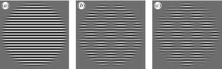
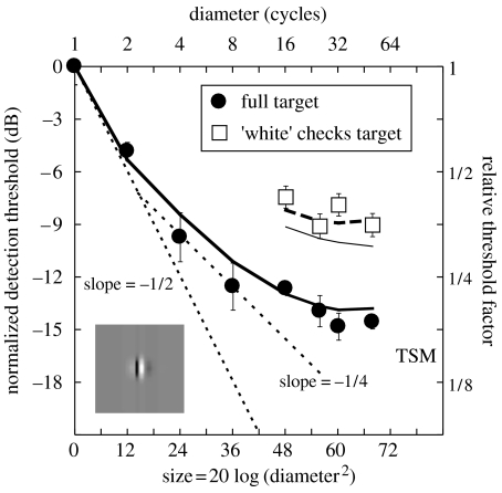
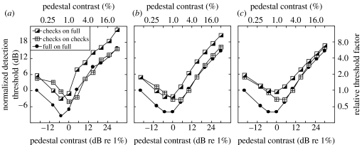
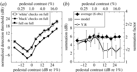
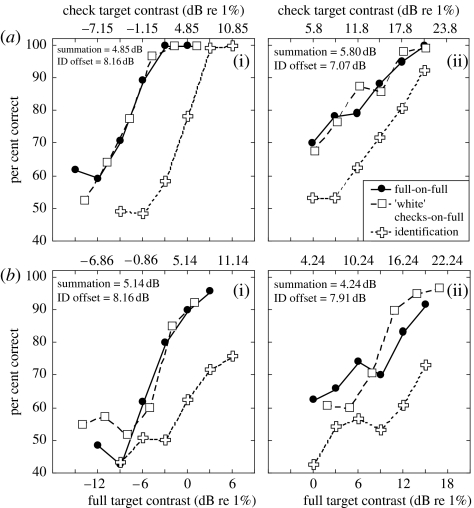
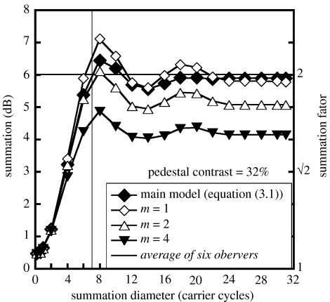
The initial image-processing stages of visual cortex are well suited to a local (patchwise) analysis of the viewed scene. But the world's structures extend over space as textures and surfaces, suggesting the need for spatial integration. Most models of contrast vision fall shy of this process because (i) the weak area summation at detection threshold is attributed to probability summation (PS) and (ii) there is little or no advantage of area well above threshold. Both of these views are challenged here. First, it is shown that results at threshold are consistent with linear summation of contrast following retinal inhomogeneity, spatial filtering, nonlinear contrast transduction and multiple sources of additive Gaussian noise. We suggest that the suprathreshold loss of the area advantage in previous studies is due to a concomitant increase in suppression from the pedestal. To overcome this confound, a novel stimulus class is designed where: (i) the observer operates on a constant retinal area, (ii) the target area is controlled within this summation field, and (iii) the pedestal is fixed in size. Using this arrangement, substantial summation is found along the entire masking function, including the region of facilitation. Our analysis shows that PS and uncertainty cannot account for the results, and that suprathreshold summation of contrast extends over at least seven target cycles of grating.
Figures
Similar articles
-
Contrast integration over area is extensive: a three-stage model of spatial summation.J Vis. 2011 Dec 16;11(14):14. doi: 10.1167/11.14.14. J Vis. 2011. PMID: 22178702
-
Spatially extensive summation of contrast energy is revealed by contrast detection of micro-pattern textures.J Vis. 2010 Jul 1;10(8):14. doi: 10.1167/10.8.14. J Vis. 2010. PMID: 20884589
-
A cortical pooling model of spatial summation for perimetric stimuli.J Vis. 2006 Oct 13;6(11):1159-71. doi: 10.1167/6.11.2. J Vis. 2006. PMID: 17209726 Free PMC article.
-
Mechanisms for spatial integration in visual detection: a model based on lateral interactions.Spat Vis. 1999;12(2):187-209. doi: 10.1163/156856899x00111. Spat Vis. 1999. PMID: 10221427 Review.
-
Modelling spatial vision at the threshold level.Spat Vis. 1992;6(1):25-60. doi: 10.1163/156856892x00046. Spat Vis. 1992. PMID: 1536828 Review.
Cited by
-
Adaptation to implied tilt: extensive spatial extrapolation of orientation gradients.Front Psychol. 2013 Jul 19;4:438. doi: 10.3389/fpsyg.2013.00438. eCollection 2013. Front Psychol. 2013. PMID: 23882243 Free PMC article.
-
Suprathreshold length summation.J Vis. 2023 Jul 3;23(7):17. doi: 10.1167/jov.23.7.17. J Vis. 2023. PMID: 37505916 Free PMC article.
-
The Effects of tDCS Across the Spatial Frequencies and Orientations that Comprise the Contrast Sensitivity Function.Front Psychol. 2015 Nov 27;6:1784. doi: 10.3389/fpsyg.2015.01784. eCollection 2015. Front Psychol. 2015. PMID: 26640448 Free PMC article.
-
Spatio-chromatic contrast sensitivity under mesopic and photopic light levels.J Vis. 2020 Apr 9;20(4):23. doi: 10.1167/jov.20.4.23. J Vis. 2020. PMID: 32347909 Free PMC article.
-
Evidence for an Optimal Algorithm Underlying Signal Combination in Human Visual Cortex.Cereb Cortex. 2017 Jan 1;27(1):254-264. doi: 10.1093/cercor/bhw395. Cereb Cortex. 2017. PMID: 28031176 Free PMC article.
References
Publication types
MeSH terms
Grants and funding
LinkOut - more resources
Full Text Sources
