. 2007 Dec;17(12):3179-88.
doi: 10.1007/s00330-007-0724-9. Epub 2007 Sep 13.

Dual-source computed tomography in patients with acute chest pain: feasibility and image quality

Affiliations

Dual-source computed tomography in patients with acute chest pain: feasibility and image quality

Thomas Schertler et al. Eur Radiol. 2007 Dec.

Abstract

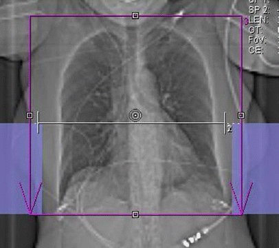
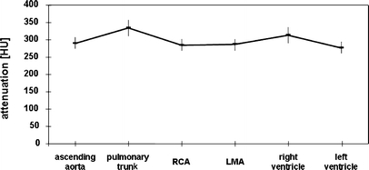
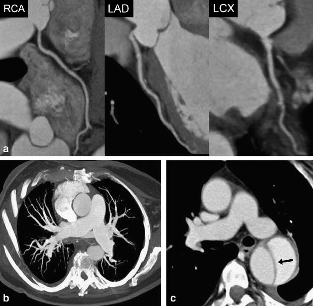
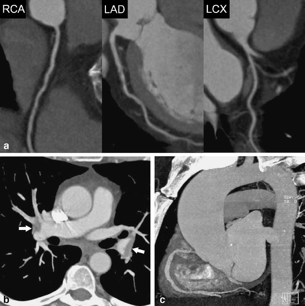
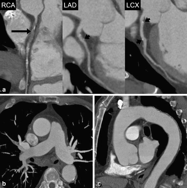
The aim of this study was to determine the feasibility and image quality of dual-source computed tomography angiography (DSCTA) in patients with acute chest pain for the assessment of the lung, thoracic aorta, and for pulmonary and coronary arteries. Sixty consecutive patients (32 female, 28 male, mean age 58.1+/-16.3 years) with acute chest pain underwent contrast-enhanced electrocardiography-gated DSCTA without prior beta-blocker administration. Vessel attenuation of different thoracic vascular territories was measured, and image quality was semi-quantitatively analyzed by two independent readers. Image quality of the thoracic aorta was diagnostic in all 60 patients, image quality of pulmonary arteries was diagnostic in 59, and image quality of coronary arteries was diagnostic in 58 patients. Pairwise intraindividual comparisons of attenuation values were small and ranged between 1+/-6 HU comparing right and left coronary artery and 56+/-9 HU comparing the pulmonary trunk and left ventricle. Mean attenuation was 291+/-65 HU in the ascending aorta, 334+/-93 HU in the pulmonary trunk, and 285+/-66 HU and 268+/-67 HU in the right and left coronary artery, respectively. DSCTA is feasible and provides diagnostic image quality of the thoracic aorta, pulmonary and coronary arteries in patients with acute chest pain.


Figures

Fig. 1
Fig. 1
Scan topogram illustrating planning of the chest pain protocol. The scan range covered the entire chest (red box). Premonitoring for bolus tracking was performed at the level of the aortic root (white line). The border for full tube current for the heart and half tube current for the upper lung is set approximately 2 cm below the tracheal bifurcation. It is delimited by a virtual horizontal line connecting the upper ends of the blue boxes on both sides
Fig. 2
Fig. 2
Mean attenuation values (HU) within different vessels indicating a relatively homogenous contrast distribution between the different thoracic vascular territories. RCA = right coronary artery; LMA = left main coronary artery
Fig. 3
Fig. 3
A 63-year-old female patient admitted to the emergency department with acute chest pain. (a) Curved multiplanar reformations along the centerline of the right coronary (RCA), left anterior descending (LAD), and the left circumflex artery (RCX) allow excluding significant coronary stenosis in this patient. Mean heart rate during DSCTA was 71 bpm. (b) Thin-slab transverse maximum intensity projection shows no evidence of pulmonary embolism. (c) Transverse image at the level of the pulmonary trunk demonstrates acute aortic dissection type B (arrow) with mild left-sided pleural effusion
Fig. 4
Fig. 4
A 58-year-old female patient admitted to the emergency department with acute chest pain. (a) Curved multiplanar reformations along the centerline of the right coronary (RCA), left anterior descending (LAD), and the left circumflex artery (LCX) demonstrate normal coronary arteries and no evidence of stenosis. Mean heart rate during DSCTA was 63 bpm. (b) Thin-slab transverse maximum intensity projection show bilateral pulmonary embolism (arrows). (c) Oblique-sagittal thin-slab maximum intensity projection demonstrates the thoracic aorta without evidence of disease
Fig. 5
Fig. 5
A 71-year-old male patient admitted to the emergency department with acute chest pain. (a) Curved multiplanar reformations along the centerline of the right coronary (RCA), left anterior descending (LAD), and the left circumflex artery (LCX) show occlusion of the proximal RCA (long arrow) and vessel wall calcifications without significant stenosis in the proximal and middle segment of the LAD and LCX (short arrows). Mean heart rate during DSCTA was 73 bpm. (b) Thin-slab transverse maximum intensity projection demonstrates normal opacification of pulmonary arteries with no evidence of embolism. (c) Oblique-sagittal thin-slab maximum intensity projection demonstrates the thoracic aorta with minimal atherosclerotic wall changes, but with no evidence of potential causes for acute chest pain
Fig. 6
Fig. 6
Flow chart of a generally accepted clinical pathway for patients with cardiac and non-cardiac causes of acute chest pain. CT represents the standard of reference in patients with suspicion of acute aortic syndromes or pulmonary embolism (light gray boxes). The added value of CT in the evaluation of acute coronary syndrome has been already demonstrated [8] (moderate gray box). The potential future role of CT in patients with acute chest pain might be at an even earlier point of diagnostic work-up (dark gray boxes) to rule out life-threatening coronary, pulmonary, and aortic disease and to guide adequate therapeutic interventions

References

    1. {'text': '', 'ref_index': 1, 'ids': [{'type': 'DOI', 'value': '10.1001/jama.294.20.2623', 'is_inner': False, 'url': 'https://doi.org/10.1001/jama.294.20.2623'}, {'type': 'PubMed', 'value': '16304077', 'is_inner': True, 'url': 'https://pubmed.ncbi.nlm.nih.gov/16304077/'}]}
    2. Swap CJ, Nagurney JT (2005) Value and limitations of chest pain history in the evaluation of patients with suspected acute coronary syndromes. Jama 294:2623–2629 - PubMed
    1. {'text': '', 'ref_index': 1, 'ids': [{'type': 'PubMed', 'value': '10190875', 'is_inner': True, 'url': 'https://pubmed.ncbi.nlm.nih.gov/10190875/'}]}
    2. Zimmerman J et al (1999) Diagnostic marker cooperative study for the diagnosis of myocardial infarction. Circulation 99:1671–1677 - PubMed
    1. {'text': '', 'ref_index': 1, 'ids': [{'type': 'DOI', 'value': '10.1067/mem.2002.129506', 'is_inner': False, 'url': 'https://doi.org/10.1067/mem.2002.129506'}, {'type': 'PubMed', 'value': '12447334', 'is_inner': True, 'url': 'https://pubmed.ncbi.nlm.nih.gov/12447334/'}]}
    2. Fesmire FM et al (2002) The Erlanger chest pain evaluation protocol: a one-year experience with serial 12-lead ECG monitoring, two-hour delta serum marker measurements, and selective nuclear stress testing to identify and exclude acute coronary syndromes. Ann Emerg Med 40:584–594 - PubMed
    1. {'text': '', 'ref_index': 1, 'ids': [{'type': 'DOI', 'value': '10.2214/AJR.05.2240', 'is_inner': False, 'url': 'https://doi.org/10.2214/ajr.05.2240'}, {'type': 'PubMed', 'value': '17056911', 'is_inner': True, 'url': 'https://pubmed.ncbi.nlm.nih.gov/17056911/'}]}
    2. Hoffmann U et al (2006) MDCT in early triage of patients with acute chest pain. AJR Am J Roentgenol 187:1240–1247 - PubMed
    1. {'text': '', 'ref_index': 1, 'ids': [{'type': 'DOI', 'value': '10.1253/circj.69.1047', 'is_inner': False, 'url': 'https://doi.org/10.1253/circj.69.1047'}, {'type': 'PubMed', 'value': '16127184', 'is_inner': True, 'url': 'https://pubmed.ncbi.nlm.nih.gov/16127184/'}]}
    2. Sato Y et al (2005) Efficacy of multislice computed tomography for the detection of acute coronary syndrome in the emergency department. Circ J 69:1047–1051 - PubMed

Publication types